Armed conflict and humanitarian crises
White phosphorus: a chemical burn that reignites
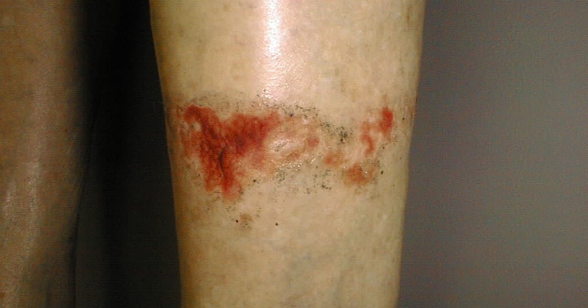



Table of contents
- What makes white phosphorus different
- How the burn behaves on the skin
- Systemic toxicity: calcium, heart, liver and kidney
- First response: extinguish without spreading
- What the evidence says about copper sulfate
- Civilian and military scenarios and the differential
- Studies at a glance
- How NEXO helps train this reasoning
- Frequently asked questions
- Take-home messages
- References
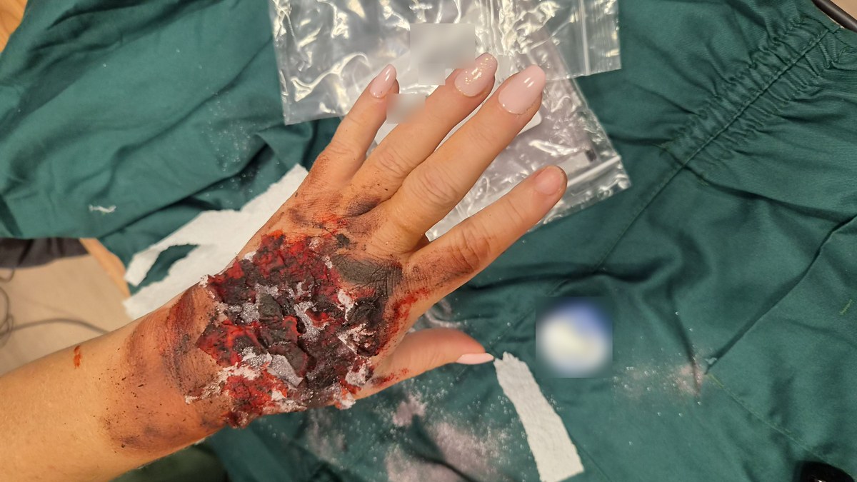
In a fictional case, an adult arrives at the emergency department with the hand and forearm covered by dry gauze. The skin has erythematous areas with adherent dark spots. On the way, the team saw smoke coming from the dressing when it was opened to air. The clinical question is not only "thermal or chemical burn?": it is whether there is still active white phosphorus (WP), whether the wound is wet enough to prevent re-ignition, and whether there is already a sign of systemic toxicity.
White phosphorus is an agent that burns on contact with oxygen, produces extreme heat and leaves particles that can re-ignite when the wound dries. The full-text literature describes deep skin injury, risk of arrhythmia from hypocalcemia even in a small body area, and hepatic and renal failure. This text separates what is mechanism, what is immediate management and what is systemic surveillance. Airway management for carbon monoxide and cyanide smoke is in the post on burns and smoke; that for nerve agents and organophosphates, in the one on cholinergic syndrome. Here the axis is different: keep it wet, find the particle and monitor calcium.
The joint reading of Wang (review), Cochrane (copper sulfate) and the case reports by Aviv, Phoenix and Ng (small areas with active management) forms the core of this post. Witkowski adds the experimental comparison of dressings; Shingleton, the austere view; Giurgiu and Żwierełło, the place of chemical burns in the set of admissions and in the general classification (Wang et al., 2025; Barqouni et al., 2014; Aviv et al., 2017).
Key points
- WP has a melting point near 44.1 °C and can ignite spontaneously near 34 to 40 °C; combustion exceeds 1,300 °C (Wang et al., 2025).
- Even with a burned area smaller than 10 percent of body surface, there are reports of fatal arrhythmia attributed to acute hypocalcemia (Wang et al., 2025).
- The frontline is to exclude oxygen (immersion or dressing saturated with cold water), remove particles under ultraviolet light or a Wood lamp when available, and avoid copper sulfate because of nephrotoxicity (Wang et al., 2025; Barqouni et al., 2014).
- In an experimental bacon model, wet gauze covering the entire area for about 3 minutes was the most effective option among the dressings tested to extinguish and remove pieces of WP (Witkowski et al., 2015).
- In the case reports by Aviv (about 2 percent TBSA) and Phoenix (less than 1 percent TBSA), small areas still required careful debridement; in both patients electrolytes did not change, but the authors recall the risk of hypocalcemia (Aviv et al., 2017; Phoenix et al., 2024).
What makes white phosphorus different
WP is an allotrope of phosphorus that oxidizes easily in air. The review by Wang and colleagues summarizes central thermochemical properties: melting point of 44.1 °C, autoignition around 34 to 40 °C under ambient conditions and combustion temperatures above 1,300 °C (Wang et al., 2025). In another passage, the same review cites an ignition point near 30 °C, which reinforces the idea of spontaneous combustion at room temperature (Wang et al., 2025).
In clinical practice this matters for three reasons. First, the injury is mixed: thermal from the temperature and chemical from the combustion products and the phosphoric acid generated. Second, unburned particles can remain in the wound and re-ignite when the dressing dries or when the patient is moved to a dry environment (Wang et al., 2025; Aviv et al., 2017). Third, systemic absorption of phosphorus and the drop in serum calcium can occur early, including with a small burned surface (Wang et al., 2025).
The general classification of chemical burns places this group at about 3 percent of all burns in pathophysiology reviews (Żwierełło et al., 2023). In a burn center, an eight-year series found 33 patients with chemical burn, equivalent to 4.39 percent of burn admissions in that service (Giurgiu et al., 2025). These numbers describe the set of chemical burns, not only WP; even so, they place the problem as relatively rare in everyday civilian life and potentially critical when it appears in conflict or industrial accident.
The density cited by Wang is 1.82 g/cm³, with a boiling point of 280 °C (Wang et al., 2025). These numbers help to understand why the material liquefies in warm water and why the team must insist on cold water. Combustion smoke also irritates the airways; even so, the focus of this text remains the skin particle and systemic toxicity, leaving the detailed management of carbon monoxide and cyanide for the neighboring post on burns and smoke.
The contrast with the classic thermal burn and with smoke inhalation is clinical: there the immediate risk is usually the airway, carbon monoxide and cyanide (burns and smoke; smoke in the cabin). In WP, the additional immediate risk is the particle that is still burning and hypocalcemia.
How the burn behaves on the skin
Skin hit by WP may show erythema, necrosis, dark particle spots and the characteristic odor described in the series. In reports with full text, depth ranges from deep dermal to full thickness even in small areas. Aviv and colleagues described a 40-year-old woman with about 2 percent TBSA: 0.5 percent on the hands (dorsum and palm), 1 percent on the right medial calf and 0.5 percent on the dorsum of the right foot (Aviv et al., 2017). Phoenix and colleagues described a 27-year-old fisherman with a chemical burn of the right hand of less than 1 percent TBSA after contact with a substance suspected to be WP on board (Phoenix et al., 2024). Ng and colleagues reported a 22-year-old soldier with a phosphorus burn on the back after improper handling of grenades (Ng et al., 2015).
The practical message from the reports is the same: a small area does not mean a trivial injury. The embedded particle continues local damage and may migrate or reignite. In an austere environment, military burn wound care guidance reinforces immersion in cold water and the aid of a Wood lamp to find fragments (Shingleton et al., 2024).
Giurgiu and colleagues, when analyzing the face and neck subgroup (15 of 33 patients with chemical burn), reinforce that cervicofacial topography adds challenges of edema, airway and scarring, even when the chemical cause is not WP (Giurgiu et al., 2025). For the clinician attending conflict or accident, the cross-cutting lesson is to examine face, hands and scalp with the same attention given to the trunk: small particles hide in folds and under crusts.
In conflict, the child and adult injured by explosion may have a burn associated with blast and combined injury; the priority reasoning (hemorrhage, airway, still active particles) intersects with child injured in war, blast trauma and field hemorrhage control. WP adds the rule of not letting the wound dry.
Systemic toxicity: calcium, heart, liver and kidney
Wang's review highlights arrhythmia-induced hypocalcemia, acute respiratory distress syndrome and hepatorenal failure as axes of the systemic pathophysiology of WP (Wang et al., 2025). The same review associates deaths from ventricular arrhythmia with burns of less than 10 percent TBSA, attributed to acute hypocalcemia with serum calcium (Ca2+) below 1.8 mmol/L (Wang et al., 2025). Aviv summarizes serious physiological changes, including hypocalcemia and hyperphosphatemia with calcium-phosphate shift, described as early as 1 hour after the burn (Aviv et al., 2017). Phoenix cites hypocalcemia and hypophosphatemia with potential sequelae of arrhythmia, in addition to hepatic and renal toxicity (Phoenix et al., 2024).
At the bedside, this translates into three actions (our synthesis): measure calcium and phosphorus early, monitor the electrocardiogram and do not discharge the patient just because the area looks small. Ng recalls the correction of hypocalcemia with intravenous calcium gluconate in the context of the management described in the series (Ng et al., 2015). Renal surveillance articulates with the reasoning of acute kidney injury and, in crush disasters, with crush syndrome, without mixing the mechanisms.
Our synthesis: WP calls for a laboratory and monitoring package that an isolated thermal burn of the same area often would not require right away. The focus is calcium and rhythm, not just the wound.
First response: extinguish without spreading
Wang lists three imperatives of the emergency protocol: immediate exclusion of oxygen by immersion or wet dressing; mechanical debridement in preference to copper sulfate; and continuous cardiac monitoring to prevent arrhythmia (Wang et al., 2025). The correction published in the same journal corrects only one author affiliation and does not change these clinical axes (Wang et al., 2025 correction).
During transport, the gauze must stay saturated. Wang mentions hydrogel-type dressings when keeping the gauze wet is difficult (Wang et al., 2025). The exact choice of product depends on what the service has validated; the principle is to prevent re-exposure to air until complete debridement.
The sequence below is our synthesis from the sources with full text:
- Remove contaminated clothing carefully so as not to spread particles.
- Exclude oxygen from the area: immersion in cold water (Wang cites water below 25 °C) or water-saturated dressings (Wang et al., 2025).
- Change saturated dressings frequently; Wang mentions changing every 5 to 7 minutes (Wang et al., 2025).
- Avoid hot water: the melting point near 44 °C makes phosphorus liquefy and increases penetration (Wang et al., 2025).
- Store the removed particles in a container with cold water for safe disposal (Ng et al., 2015; Shingleton et al., 2024).
Witkowski and colleagues compared first aid dressings in a bacon model covered by a military uniform. All dressings tested extinguished the WP when they covered the entire burning area; wet gauze stood out for extinguishing, removing pieces and reducing immediate re-ignition when kept in place for about 3 minutes (Witkowski et al., 2015). The water jet was described as dangerous in the model because it splashed and transferred pieces of WP to other areas (Witkowski et al., 2015). This experimental finding does not replace a clinical trial in humans, but it aligns with the clinical emphasis on continuous wet coverage instead of a strong jet.
Cochrane also mentions irrigating the wound continuously with water or saline (Barqouni et al., 2014), which is different from a strong jet. The contemporary consensus on second-degree wounds reinforces general principles of moisture and wound protection, without specifically addressing WP (Ji et al., 2024). Our synthesis for WP: wet, cold, full coverage, no jet that spreads.
On a scene with multiple casualties, triage priority follows START and SALT and the service's capacity, as in surge capacity. Radioactive decontamination of a dirty bomb and the combined injury of radiation, trauma and burn are neighboring axes: here the active "contamination" is the WP particle that still reacts with the air.
Finding and removing the particles
After extinguishing the flame, the decisive step is to find what has not yet burned. Wang describes mechanical extraction with blunt forceps under a Wood lamp, which makes WP fluoresce; in its absence, ultraviolet flashlights or fluorescent detection devices (Wang et al., 2025). Aviv also points to the Wood lamp to identify residual luminescent particles with low additional risk (Aviv et al., 2017). Ng mentions copper sulfate or ultraviolet light for identification, and reinforces immersion in cold water during debridement to prevent new ignition (Ng et al., 2015). Shingleton, in the austere context, cites the Wood lamp to find fragments after immersion (Shingleton et al., 2024).
The practical order below is our synthesis from the sources:
- Keep the area moist at all times.
- Examine under ultraviolet light or a Wood lamp, or in a dark room observing chemiluminescence.
- Remove visible particles with a blunt instrument.
- Place the removed material in a jar with cold water.
- Repeat the examination until no fluorescence remains.
- Only then move on to the definitive dressing and depth assessment.
Incomplete removal explains the re-ignition during transport and in the ward. Aviv warns that aggressive irrigation with a lot of water can carry particles to previously intact skin, where they re-ignite on exposure to air (Aviv et al., 2017). Wang warns of the same phenomenon when the dressing dries (Wang et al., 2025).
What the evidence says about copper sulfate
Copper sulfate appears in the history of WP management because it darkens the particles and makes them easier to see. The Cochrane review by Barqouni and colleagues on interventions for phosphorus burns included two non-randomized comparative studies of patients treated with and without copper sulfate (Barqouni et al., 2014). The authors concluded that these studies provide no evidence in favor of copper sulfate; the small amount of available evidence suggests it may be harmful (Barqouni et al., 2014). The Cochrane review reinforces common-sense measures: act quickly, remove clothing, irrigate the wound continuously with water or saline and remove the particles (Barqouni et al., 2014).
Wang classifies copper sulfate as controversial and contraindicated due to the risk of hemolysis, toxicity in glucose-6-phosphate dehydrogenase deficiency and nephrotoxicity, and prioritizes mechanical debridement (Wang et al., 2025). Aviv reviews the historical evolution of copper sulfate use and highlights skin absorption and toxicity (Aviv et al., 2017). Ng still mentions copper sulfate as a historical identification option, but the contemporary line of full-text reviews points to ultraviolet light and mechanical removal (Ng et al., 2015; Wang et al., 2025).
For the clinician: if the institution still has copper sulfate in its old protocol, the current reading of open sources recommends replacing identification with ultraviolet light and removal with a mechanical technique under moisture.
Civilian and military scenarios and the differential
Giurgiu's series does not isolate WP, but it offers a picture of the relative volume of chemical burns at a referral center: 33 admissions in eight years (Giurgiu et al., 2025). This explains why many civilian clinicians have never seen a case and, at the same time, why a single poorly conducted encounter (dry dressing, copper sulfate, discharge too early without calcium) can be serious. In a military or austere setting, Shingleton and the team at the U.S. Army Institute of Surgical Research place WP among the chemical burns that require immersion and identification of fragments before the definitive dressing (Shingleton et al., 2024).
Civilian reports (a fisherman, an accident with fireworks or an agricultural product) and military ones (a grenade, ammunition) share the same local algorithm, but differ in the context of blast, multiple casualties and time to the hospital. Phoenix proposes an algorithm for a civilian hand burn suspected of WP, emphasizing washing, identification and dressing (Phoenix et al., 2024). Wang discusses the humanitarian challenge of WP munitions and the need for clear protocols in emergency care (Wang et al., 2025).
The differential in the emergency department includes:
- Pure thermal burn and smoke inhalation (burns and smoke).
- Other chemical burns (acids, alkalis, hydrofluoric acid): different mechanism and antidotes; HF must not be confused with WP.
- Nerve agents and organophosphates (cholinergic syndrome).
- Blast injury with associated burn (blast trauma).
When the presentation mixes blast and burn, the reasoning of blast trauma and of the child wounded in war organizes life priorities. WP enters as a wound modifier: after hemorrhage and the airway, the question "can this burn still re-ignite?" changes the dressing and the laboratory work. In injury combined with radiation, the post on radiation, trauma and burns covers another axis; here the residual "activity" is chemical, not radioactive.
The question that changes management: is the wound still smoking or glowing in the dark? Is there a dry dressing? Is there paresthesia, tetany, arrhythmia or a laboratory calcium abnormality? If so, treat as active WP until proven otherwise.
Studies at a glance
| Source | Design / setting | Key finding used in this text |
|---|---|---|
| Wang 2025 | Narrative review (Front Public Health) | Mechanism, 44.1 °C fusion, hypocalcemia with TBSA under 10 percent (Wang et al., 2025) |
| Aviv 2017 | Single-patient report and review (Sheba) | Woman, about 2 percent TBSA; Wood; normal electrolytes in the case, risk described in the review |
| Phoenix 2024 | Civilian case report (hand) | Fisherman, under 1 percent TBSA; normal ECG and electrolytes; hand algorithm |
| Ng 2015 | Single-patient report (Johns Hopkins) | Soldier, back; irrigation; calcium gluconate |
| Witkowski 2015 | Experiment in a bacon model | In the bacon model: wet gauze about 3 minutes; a jet spreads particles |
| Barqouni 2014 | Cochrane (2 non-randomized comparative studies) | No evidence in favor of copper sulfate; possible harm (Barqouni et al., 2014) |
| Giurgiu 2025 | Single-center series (8 years) | 33 patients with chemical burns; 4.39 percent of burn admissions at the center (Giurgiu et al., 2025) |
| Shingleton 2024 | Austerity care review | Wood and immersion for WP fragments |
| Żwierełło 2023 | Pathophysiology review | Chemical burns about 3 percent of all burns |
| Ji 2024 | Second-degree wound consensus | General principles of wound moisture |
Sources: Wang et al., 2025; Aviv et al., 2017; Phoenix et al., 2024; Ng et al., 2015; Witkowski et al., 2015; Barqouni et al., 2014; Giurgiu et al., 2025; Shingleton et al., 2024; Żwierełło et al., 2023; Ji et al., 2024.
How NEXO helps train this reasoning
In NEXO, the case of the day is fictional, with a real clinical presentation and up to six progressive clues, and you propose the diagnosis by searching for the ICD-10 code. One clue may bring "wound that smokes again when it dries" or "low calcium with a small area"; another may show positive ultraviolet light on the skin. The five-level proximity feedback helps separate thermal burn, other chemical burn and WP without turning the training into a field protocol checklist. The useful exercise is to say out loud, at each clue, whether the priority now is moisture, particle removal or labs and ECG.
Frequently asked questions
Why does the wound reignite?
Because unconsumed WP particles oxidize again when they contact the air, especially if the dressing dries (Wang et al., 2025; Aviv et al., 2017).
Can I dry the wound to photograph it or transfer the patient?
The logic of the sources is to keep moisture until the particles are completely removed and to transport under a saturated dressing (Wang et al., 2025; Witkowski et al., 2015). Drying for documentation increases the risk of reignition.
Is copper sulfate still part of the protocol?
The Cochrane review found no evidence in favor and pointed to a risk of harm; Wang prioritizes mechanical debridement and ultraviolet light (Barqouni et al., 2014; Wang et al., 2025).
Does a small area rule out blood tests and ECG?
Wang links fatal arrhythmia to hypocalcemia even with TBSA under 10 percent. In the Aviv (about 2 percent TBSA) and Phoenix (under 1 percent TBSA) cases the electrolytes were normal, but both reports describe the risk of hypocalcemia and arrhythmia, and Phoenix did an ECG and electrolytes on admission (Wang et al., 2025; Aviv et al., 2017; Phoenix et al., 2024).
Does a strong water jet help?
In the Witkowski experimental model, the jet spread pieces of WP; full wet coverage was preferable (Witkowski et al., 2015). Controlled continuous irrigation and immersion differ from a turbulent jet.
Take-home messages
- Treat WP as a chemical burn that can still burn: keep the area wet and cool until the particles are removed.
- Look for fluorescence (Wood lamp or ultraviolet) and remove mechanically; store the material in cold water.
- Set copper sulfate aside: Cochrane does not support it and recent reviews point to toxicity.
- Order calcium, phosphorus and ECG even with a small area; hypocalcemia is the thread linking wound and arrhythmia.
- Mentally separate WP from thermal burn with smoke and from organophosphate: the blog's neighboring articles cover those axes without repeating this management.
References
- Wang M, Bai Y, Zhang X, Zhang J, Kang A. White phosphorus munitions: pathophysiology, clinical management, and multidisciplinary perspectives on burn injuries and humanitarian challenges. Front Public Health. 2025;13:1632840. PMID: [40786173](https://pubmed.ncbi.nlm.nih.gov/40786173/). PMC: [PMC12331754](https://www.ncbi.nlm.nih.gov/pmc/articles/PMC12331754/). Correction: Front Public Health. 2025;13:1673762. PMID: [40860547](https://pubmed.ncbi.nlm.nih.gov/40860547/). PMC: [PMC12371926](https://www.ncbi.nlm.nih.gov/pmc/articles/PMC12371926/).
- Aviv U, Kornhaber R, Harats M, Haik J. The burning issue of white phosphorus: a case report and review of the literature. Disaster Mil Med. 2017;3:6. PMID: [28861275](https://pubmed.ncbi.nlm.nih.gov/28861275/). PMC: [PMC5577774](https://www.ncbi.nlm.nih.gov/pmc/articles/PMC5577774/).
- Phoenix E, Suwanwalaikorn V, Wilkinson J, Morrison CM, Dolan RT. White phosphorous civilian hand burns - An aid to timely treatment of a rare entity. Trauma Case Rep. 2024;50:100983. PMID: [38496001](https://pubmed.ncbi.nlm.nih.gov/38496001/). PMC: [PMC10940903](https://www.ncbi.nlm.nih.gov/pmc/articles/PMC10940903/).
- Ng NYB, Abdullah A, Milner SM. A phosphorus burn. Eplasty. 2015;15:ic15. PMID: [25834695](https://pubmed.ncbi.nlm.nih.gov/25834695/). PMC: [PMC4364261](https://www.ncbi.nlm.nih.gov/pmc/articles/PMC4364261/).
- Witkowski W, Surowiecka-Pastewka A, Biesaga M, Gierczak T. Experimental comparison of efficiency of first aid dressings in burning white phosphorus on bacon model. Med Sci Monit. 2015;21:2361-2366. PMID: [26264209](https://pubmed.ncbi.nlm.nih.gov/26264209/). PMC: [PMC4538885](https://www.ncbi.nlm.nih.gov/pmc/articles/PMC4538885/).
- Barqouni L, Abu Shaaban N, Elessi K. Interventions for treating phosphorus burns. Cochrane Database Syst Rev. 2014;(6):CD008805. PMID: [24896302](https://pubmed.ncbi.nlm.nih.gov/24896302/). PMC: [PMC7173745](https://www.ncbi.nlm.nih.gov/pmc/articles/PMC7173745/).
- Giurgiu RA, Dumitru CS, Grosu-Bularda A, et al. Chemical burns: pathophysiology and therapeutic protocols - do cervico-facial injuries pose specific challenges? J Med Life. 2025;18(5):440-450. PMID: [40599149](https://pubmed.ncbi.nlm.nih.gov/40599149/). PMC: [PMC12207703](https://www.ncbi.nlm.nih.gov/pmc/articles/PMC12207703/).
- Shingleton S, Folwell J, Jones I, Gleason M, Williams A. Burn wound care strategies for the battlefield and austere settings. Eur Burn J. 2024;5(1):5. PMID: [39600013](https://pubmed.ncbi.nlm.nih.gov/39600013/). PMC: [PMC11571820](https://www.ncbi.nlm.nih.gov/pmc/articles/PMC11571820/).
- Żwierełło W, Piorun K, Skórka-Majewicz M, Maruszewska A, Antoniewski J, Gutowska I. Burns: classification, pathophysiology, and treatment: a review. Int J Mol Sci. 2023;24(4):3749. PMID: [36835171](https://pubmed.ncbi.nlm.nih.gov/36835171/). PMC: [PMC9959609](https://www.ncbi.nlm.nih.gov/pmc/articles/PMC9959609/).
- Ji S, Xiao S, Xia Z; Chinese Burn Association Tissue Repair of Burns and Trauma Committee, Cross-Straits Medicine Exchange Association of China. Consensus on the treatment of second-degree burn wounds (2024 edition). Burns Trauma. 2024;12:tkad061. PMID: [38343901](https://pubmed.ncbi.nlm.nih.gov/38343901/). PMC: [PMC10858447](https://www.ncbi.nlm.nih.gov/pmc/articles/PMC10858447/).