Long-term follow-up after radiation exposure
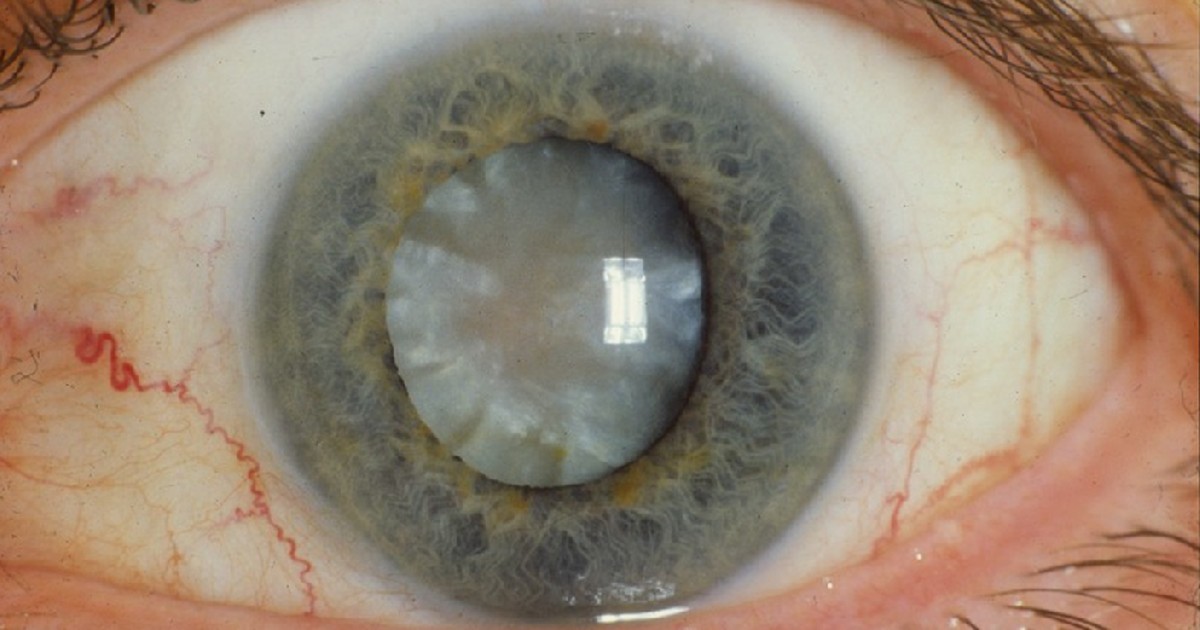
Table of contents
A 58-year-old woman comes to a routine appointment with a folder of old test results. At age 22, she lived near a scrapyard where a radioactive source was opened, had hair loss and a wound on her hand that left a scar, and chromosome analysis estimated a whole-body dose of about 0.5 Gy. She has not been to the follow-up service for years. Now she wants to know whether she is still at risk of cancer, whether the high blood pressure that has just appeared is related to the accident, whether the blurred vision is radiation cataract and whether her grandson, the son of the daughter who was born after the accident, could have inherited a problem. The family doctor has never treated anyone exposed. The scene is fictional and was written for teaching.
This text uses the case as a setting for clinical reasoning for students and health professionals. The topic is the long-term follow-up of people who received moderate or high doses of ionizing radiation, in an accident, a conflict or a treatment. The focus is what the doctor decides: what to screen for, what not to attribute to radiation, how to talk about risk and how to answer questions about children and grandchildren. It does not replace the reference service protocol for exposed people or the specialist's assessment. If you play the case of the day in NEXO, you will recognize the challenge: deciding with clues that arrive in sequence.
Key points
- The risk of solid tumors in Hiroshima and Nagasaki survivors was still higher 64 years after exposure.
- The excess of leukemia appeared first and persisted, especially acute myeloid leukemia, up to 55 years later.
- Doses above 0.5 Gy were associated with more stroke and heart disease, and the risk at lower doses is uncertain.
- The ICRP treats cataract and circulatory disease as tissue reactions with a threshold of about 0.5 Gy.
- Studies of survivors' children have so far shown no higher risk from parental exposure.
- In Goiânia, follow-up divided exposed people into groups with appointments every six months or every year.
Why this is a reasoning scenario
The appointment looks simple, but it has two opposite traps. The first is attributing everything to radiation: the high blood pressure, the cataract, any nodule and the fear for the grandson. The second is ignoring the history, treating the patient like anyone else of the same age and missing a screening that would make sense. Clinical reasoning lies in using what the long-term cohorts show to separate what is probably related to the dose, what is uncertainly related and what has no known relation.
Kamiya and colleagues summarize eight decades of studies with survivors of the atomic bombs and their children and record an excess of leukemia, of solid tumors as a whole and of most cancer sites (Kamiya and colleagues, 2025). They also record possible excesses of noncancer diseases, such as cataract and circulatory diseases (Kamiya and colleagues, 2025). In survivors' children, studies have so far shown no higher risk from parental exposure, neither at the molecular level nor at the disease level (Kamiya and colleagues, 2025).
The post on acute radiation syndrome and lymphocyte triage deals with the acute phase, and the post on accidents with radioactive sources shows how these accidents happen. This text begins when the acute phase is over and the person returns to ordinary life.
What the scene teaches
The scene trains six habits. The first is to recover the best dose estimate and the age at exposure. The second is to know which cancers have a clearer relation to radiation. The third is to treat common cardiovascular risk factors with the same care as always. The fourth is to examine the lens with a slit lamp. The fifth is to answer questions about children with what the data show. The sixth is to reconnect the person with the follow-up service.
What is not at stake
The text does not cover the acute phase, recent skin injuries or decontamination. It also does not cover the risk of low doses from imaging exams, which is a matter of patient radiation protection. The focus is the person who received a moderate or high dose and wants to know what to do in the following decades.
What the large cohorts show
Almost everything known about late effects comes from the survivors of Hiroshima and Nagasaki. Ozasa and colleagues describe that the Life Span Study has about 120,000 people followed since 1950 (Ozasa and colleagues, 2018). The cohorts of those exposed in utero and of children of survivors have about 3,600 and 77,000 people, followed since 1945 (Ozasa and colleagues, 2018). Each person's dose was estimated from their position at the moment of the explosion and from shielding conditions (Ozasa and colleagues, 2018).
Outcomes include vital status, cause of death and cancer incidence (Ozasa and colleagues, 2018). Subcohorts were invited for periodic health examinations and to donate biological samples (Ozasa and colleagues, 2018). Wakeford and Laurier note that the risk estimates from these cohorts were used by the ICRP as the main technical basis for its recommendations, from 1965 through the 2007 recommendations (Wakeford and Laurier, 2025). They also record that, 80 years after the explosions, the survivors still provide information about risks decades after an exposure at a young age (Wakeford and Laurier, 2025).
Why this matters for the patient
Cohorts measure risk in groups, not the fate of one person. The patient in the scene has an estimated dose, an age at exposure and a sex, and these three data points change the risk. Kamiya and colleagues record that, for solid tumors as a whole, exposure at a younger age, a lower attained age and female sex are associated with greater radiation sensitivity, but results vary by cancer site (Kamiya and colleagues, 2025). The physician uses these data to choose what to look at more closely, not to predict the disease.
The limits of the comparisons also need to be kept in mind. An explosion delivers an acute whole-body dose, while an orphan source usually delivers a prolonged and uneven dose, with part of the body much more irradiated. The patient's estimate from chromosomes is the best information available, but it carries uncertainty.
Cancer after exposure
Cancer is the patient's main concern and the most studied late effect. The data show a different time pattern for leukemia and for solid tumors.
Leukemia
Hsu and colleagues record that the sharp increase in leukemia risk was the first late effect and the most striking one seen in the survivors (Hsu and colleagues, 2013). The analysis included 113,011 people from the cohort with 3.6 million person-years of follow-up, from 1950 through the end of 2001 (Hsu and colleagues, 2013). Two to three years after the explosions, physicians in Hiroshima and Nagasaki noticed a much higher rate of leukemia in children who lived near the hypocenter (Hsu and colleagues, 2013).
The excess of leukemia, in general, decreased with attained age and with time since exposure, but the radiation-related excess, especially of acute myeloid leukemia, persisted throughout the follow-up, up to 55 years later (Hsu and colleagues, 2013). Even in the last 12 years of follow-up, 45 to 55 years after exposure, the radiation-related leukemia risk at 1 Gy was estimated at twice the baseline risk (Hsu and colleagues, 2013). Chronic lymphocytic leukemia and adult T-cell leukemia did not appear to be radiation-related (Hsu and colleagues, 2013). The authors found no radiation-related excess of Hodgkin lymphoma or multiple myeloma (Hsu and colleagues, 2013).
Solid tumors
Brenner and colleagues identified 22,538 solid tumors in 105,444 people from 1958 to 2009 (Brenner and colleagues, 2025). In women, the linear model best described the risk of solid tumors as a whole, with an excess relative risk of 0.64 per Gy (Brenner and colleagues, 2025). In men, the best fit was a linear-quadratic model with upward curvature, with an excess relative risk of 0.20 at 1 Gy (Brenner and colleagues, 2025). The excess relative risk decreased with attained age and with age at exposure (Brenner and colleagues, 2025). Sixty-four years after exposure, the risk of solid tumors was still higher in those exposed (Brenner and colleagues, 2025).
The same analysis identified for the first time a significant relationship between dose and prostate cancer (Brenner and colleagues, 2025). For breast and uterine corpus cancer, the risk linked to radiation increased as age at exposure approached menarche, around 15 years, and decreased at older ages (Brenner and colleagues, 2025). Kamiya and colleagues list esophagus, stomach, colon, liver, gallbladder, lung, bladder, non-melanoma skin, central nervous system, thyroid and breast among the sites with a significant association (Kamiya and colleagues, 2025). They also note that the risk of thyroid cancer is strongly influenced by age at exposure, higher in the youngest, while the risk of lung cancer linked to radiation increases with age at exposure (Kamiya and colleagues, 2025).


Sarcomas
Berrington de Gonzalez and colleagues report clear evidence of more bone and soft tissue sarcomas after fractionated high-dose radiation in childhood, from 10 Gy (Berrington de Gonzalez and colleagues, 2012). Follow-up data from survivors suggested for the first time that sarcomas can also be induced by lower acute doses, below 5 Gy, at any age (Berrington de Gonzalez and colleagues, 2012). Even so, at this dose level, a radiation-linked sarcoma would be a very rare event (Berrington de Gonzalez and colleagues, 2012).
In practice, what the doctor does with these data is keep the screening recommended for age and sex, without delay, and carefully investigate warning signs. The signs that deserve prompt investigation in an exposed person are these:
- Blood count with cytopenia, blasts or unexplained leukocytosis.
- Palpable thyroid nodule or finding on imaging.
- New breast nodule, especially in those exposed in childhood or adolescence.
- Skin lesion that changes or does not heal, including in the area of old radiodermatitis.
- Soft tissue mass or persistent bone pain in the most irradiated region.
- Persistent cough, blood in the stool or unexplained weight loss.
Heart, vessels and eyes
The patient's high blood pressure raises the question about the heart. The answer requires separating what is known for high doses from what is still uncertain for lower doses.
Stroke and heart disease
Shimizu and colleagues followed 86,611 people from the Life Span Study for more than 50 years (Shimizu and colleagues, 2010). About 9,600 died of stroke and 8,400 of heart disease between 1950 and 2003 (Shimizu and colleagues, 2010). The excess relative risk was 9 percent per Gy for stroke and 14 percent per Gy for heart disease (Shimizu and colleagues, 2010). In the 0 to 0.5 Gy range, the dose-response relationship was not significant (Shimizu and colleagues, 2010). The authors conclude that doses above 0.5 Gy are associated with more risk of stroke and heart disease, but the degree of risk at lower doses is not clear (Shimizu and colleagues, 2010). Smoking, alcohol, education, occupation, obesity and diabetes hardly changed the estimates (Shimizu and colleagues, 2010).
Schöllnberger and colleagues reanalyzed the same data with several models and found sublinear curves for cerebrovascular disease and heart disease at low and moderate doses (Schöllnberger and colleagues, 2018). They note that the analysis gives no conclusive answer on the existence of risk below 0.75 Gy for cerebrovascular disease and below 2.6 Gy for heart disease (Schöllnberger and colleagues, 2018). Little and colleagues document excess risk of ischemic heart disease and stroke in groups exposed to moderate or low doses, but with patterns that are not simple and with weak evidence of non-linearity (Little and colleagues, 2021).
The 2011 ICRP statement warns that the threshold for circulatory disease may be as low as 0.5 Gy in the heart or brain (ICRP, 2011 statement). For the patient, with an estimated dose close to that value, management does not change: measure blood pressure, treat hypertension, dyslipidemia and diabetes and stop smoking, as with anyone. The dose is no reason to treat her differently, and it is also no reason to attribute the hypertension to the accident.
Lens
In April 2011, the ICRP began to consider a threshold of 0.5 Gy for effects on the lens (ICRP, 2011 statement). Little and colleagues note that high doses, above 1 Gy, are associated with posterior subcapsular cataract (Little and colleagues, 2021). They also document excess risk of posterior subcapsular and cortical cataract at lower doses and dose rates in Chernobyl liquidators, United States radiology technicians and Mayak nuclear workers, with a basically linear relationship (Little and colleagues, 2021). Nuclear cataract seems less linked to radiation (Little and colleagues, 2021). Radiation-linked glaucoma has been documented only at doses above 5 Gy (Little and colleagues, 2021).
The patient's blurred vision calls for a slit-lamp examination. The type of opacity matters: a nuclear cataract at 58 is very common and not very suggestive of radiation, while a posterior subcapsular opacity fits the history better. In any case, the indication for surgery follows visual function, as with anyone.
Other diseases
Kamiya and colleagues note, in an incidence report from 1958 to 1998, higher radiation-linked risk for cataract, uterine fibroid, thyroid disease, chronic liver disease and cirrhosis and hypertension, as well as higher prevalence of hyperparathyroidism (Kamiya and colleagues, 2025). They note that no clear associations were found between radiation and neurocognitive function in old age in those exposed in childhood or in utero (Kamiya and colleagues, 2025). The table summarizes what the data allow us to tell the patient.
| Outcome | What the data show | Source |
|---|---|---|
| Acute myeloid leukemia | Persistent excess up to 55 years later | Hsu and colleagues, 2013 |
| Solid tumors as a group | Higher risk 64 years later | Brenner and colleagues, 2025 |
| Stroke and heart disease | Association above 0.5 Gy; uncertain below | Shimizu and colleagues, 2010 |
| Posterior subcapsular cataract | Threshold considered to be 0.5 Gy | ICRP, 2011 |
| Disease in children | No higher risk observed so far | Kamiya and colleagues, 2025 |
Children and in utero exposure
The question about the grandson is the one that weighs most on the patient, and the answer from the data is reassuring, with caution. Ozasa and colleagues note that, in the children of survivors, no higher risk from parental exposure was observed for cancer or other diseases, but investigations continue because these cohorts are still relatively young (Ozasa and colleagues, 2018). Kamiya and colleagues note the same result at the molecular and disease level (Kamiya and colleagues, 2025).
In Goiânia, Costa and colleagues studied variations in DNA copy number in the children of people exposed to cesium 137 (Costa and colleagues, 2018). They report that 249 people were exposed, with absorbed doses from nearly zero up to 7 Gy (Costa and colleagues, 2018). The authors identified 473 variations in the group of children of exposed people and 238 in the control group (Costa and colleagues, 2018). The study proposes these variations as a biological marker of parental exposure, and the authors themselves note that the usefulness of this marker still needs to be investigated (Costa and colleagues, 2018). A laboratory marker is not a disease, and this finding does not change the approach to the grandson.
Exposure in utero
The patient's daughter was born after the accident, but the question comes up in other families. Sugiyama and colleagues followed 2,463 people exposed in utero to the bombs, from 1950 to 2012 (Sugiyama and colleagues, 2021). Exposure before birth was a consistent risk factor for mortality from solid tumors in women, but not in men (Sugiyama and colleagues, 2021). The effect on mortality from noncancer diseases and external causes was mediated by small head size, low birth weight and loss of parents (Sugiyama and colleagues, 2021). The post about radiation in pregnancy covers the approach during pregnancy.
These are the answers the doctor can give the family, based on the data:
- Children conceived after their parents' exposure have not so far shown more cancer or other diseases.
- Studies continue, and the absence of an effect is not definitive proof.
- The grandson should follow the usual care for his age, with no extra tests because of his grandmother.
- People exposed in utero deserve attention to growth and, in women, to cancer screening.
Organized follow-up: the Goiânia example
The Goiânia accident, in 1987, left a model of organized follow-up. Rodrigues and colleagues report that about 112,000 people were involved, of whom 249 had external or internal contamination (Rodrigues and colleagues, 2017). The state government created a care center for exposed people, with a monitoring system (Rodrigues and colleagues, 2017).
Group I, with visits every six months, included 40 people with radiodermatitis, a cytogenetic dose above 0.20 Gy or high body activity (Rodrigues and colleagues, 2017). Group II, with annual visits, included 62 people with a cytogenetic dose between 0.05 and 0.20 Gy (Rodrigues and colleagues, 2017). Group III included 880 professionals who handled contaminated material or patients (Rodrigues and colleagues, 2017). The children of the people in groups I and II were also included and are followed (Rodrigues and colleagues, 2017). The patient in the scene, with radiodermatitis and a dose of 0.5 Gy, would be in the group with visits every six months.
What the follow-up found
In the study by Rodrigues and colleagues, with 48 interviewees, the prevalence of hypertension was 25 percent, similar to that of the general population (Rodrigues and colleagues, 2017). Rahal and colleagues analyzed the incidence of breast cancer in the women of Goiânia from 1988 to 2012 (Rahal and colleagues, 2019). The incidence rose in the first 17 years and then stayed stable, but the trend suggested no association with the accident (Rahal and colleagues, 2019). Lage and colleagues studied breast cancer cases from 2001 to 2010 in the central region of the city, with seven main sources of contamination, and found no association between current radiation levels and new cases (Lage and colleagues, 2020).
These studies look at the whole city or at small groups, and they do not answer the individual question. They serve to show that organized follow-up allows the population's fear to be answered with data, and that monitoring the most exposed people remains necessary.
Risks and reasoning errors
The most common mistakes in this consultation are attributing hypertension or nuclear cataract in a 58-year-old to radiation, ordering repeat imaging without indication, which adds dose without benefit, forgetting the blood count and thyroid exam in a young exposed person, and answering questions about children with a certainty the data do not support, in either direction. The post on diagnostic uncertainty helps talk about risk without promising anything, and the post on diagnostic pause helps stop before attributing everything to the accident.
How NEXO helps train this reasoning
NEXO publishes one fictional clinical case per day, with up to six progressive clues. You can attempt the diagnosis at each clue, searching for the ICD-10 code, and you receive proximity feedback on five levels. The format trains the same skill as this scene: putting together data that arrive gradually, such as dose, age at exposure, type of cataract and common risk factors, and deciding without closing too early. The post on case-based learning explains why this format works, and the post on clinical decision-making helps choose the right exams.
Three exercises with cases
The first exercise is to redo the scene with a 70-year-old man exposed at age 10 and decide which cancer deserves more attention. The second is to redo the scene with a woman exposed at age 14 and discuss breast screening. The third is to redo the scene with a worker who took part in decontamination and received a small dose, and decide what changes compared with routine care.
Teachers and content creators can build their own cases in the space for Creators. The exercise works well in family medicine, internal medicine, oncology and occupational medicine residencies.
Frequently asked questions
For how long does the cancer risk stay higher?
In survivors, the risk of solid tumors was still higher 64 years later (Brenner et al., 2025).
Does leukemia appear early or late?
It appeared first, two to three years later, and the excess persisted for up to 55 years (Hsu et al., 2013).
Does radiation cause hypertension?
In Goiânia, prevalence in exposed people was similar to that of the general population (Rodrigues et al., 2017).
Do children of exposed people have more diseases?
So far, studies have not shown a higher risk (Kamiya et al., 2025).
What is the threshold for cataract?
The ICRP considers 0.5 Gy (ICRP, 2011 statement).
Final checklist
- I recovered the estimated dose, the method and the age at exposure.
- I kept the cancer screening recommended for age and sex, without delay.
- I ordered a blood count and examined the thyroid and the skin of the irradiated area.
- I treated blood pressure, lipids, blood glucose and smoking as I would in anyone.
- I ordered a slit-lamp exam and noted the type of opacity.
- I avoided imaging without indication.
- I answered questions about children with the data and with the uncertainty.
- I reconnected the person to the follow-up service.
Want to practice reasoning with progressive clues? Play today's case.
References
- Berrington de Gonzalez A, Kutsenko A, Rajaraman P. Sarcoma risk after radiation exposure. Clin Sarcoma Res. 2012. PMID 23036235.
- Brenner AV, Sugiyama H, Cologne J, et al. Summary of radiation effects on incidence of solid cancers in the Life Span Study of atomic bomb survivors: 1958-2009. Carcinogenesis. 2025. PMID 41144265.
- Costa EOA, Pinto IP, Gonçalves MW, et al. Small de novo CNVs as biomarkers of parental exposure to low doses of ionizing radiation of caesium-137. Sci Rep. 2018. PMID 29651024.
- Hsu WL, Preston DL, Soda M, et al. The incidence of leukemia, lymphoma and multiple myeloma among atomic bomb survivors: 1950-2001. Radiat Res. 2013. PMID 23398354.
- International Commission on Radiological Protection. Statement on Tissue Reactions. ICRP ref 4825-3093-1464. April 2011. Official source.
- Kamiya K, Sakata R, Rajaraman P. Eight decades of research on the long-term health effects of radiation in atomic bomb survivors and their offspring. Carcinogenesis. 2025. PMID 41144264.
- Lage LB, Freitas-Junior R, Corrêa RDS, et al. Evaluation of ionizing radiation as a risk factor for the incidence of breast cancer: long-term analysis after the cesium-137 accident in Goiânia, Brazil. An ecological study. Sao Paulo Med J. 2020. PMID 32813846.
- Little MP, Azizova TV, Hamada N. Low- and moderate-dose non-cancer effects of ionizing radiation in directly exposed individuals, especially circulatory and ocular diseases: a review of the epidemiology. Int J Radiat Biol. 2021. PMID 33471563.
- Ozasa K, Grant EJ, Kodama K. Japanese Legacy Cohorts: The Life Span Study Atomic Bomb Survivor Cohort and Survivors' Offspring. J Epidemiol. 2018. PMID 29553058.
- Rahal RMS, Rocha ME, Freitas-Junior R, et al. Trends in the Incidence of Breast Cancer Following the Radiological Accident in Goiânia: A 25-Year Analysis. Asian Pac J Cancer Prev. 2019. PMID 31870126.
- Rodrigues JVR, Pinto MM, Figueredo RMP, et al. Systemic Arterial Hypertension in Patients Exposed to Cesium-137 in Goiânia-GO: Prevalence Study. Arq Bras Cardiol. 2017. PMID 28562830.
- Schöllnberger H, Eidemüller M, Cullings HM, et al. Dose-responses for mortality from cerebrovascular and heart diseases in atomic bomb survivors: 1950-2003. Radiat Environ Biophys. 2018. PMID 29222678.
- Shimizu Y, Kodama K, Nishi N, et al. Radiation exposure and circulatory disease risk: Hiroshima and Nagasaki atomic bomb survivor data, 1950-2003. BMJ. 2010. PMID 20075151.
- Sugiyama H, Misumi M, Sakata R, et al. Mortality among individuals exposed to atomic bomb radiation in utero: 1950-2012. Eur J Epidemiol. 2021. PMID 33492551.
- Wakeford R, Laurier D. The importance of data from the atomic bomb survivors for international radiological protection. Carcinogenesis. 2025. PMID 41144268.