



Digital health and AIBurnout and well-being
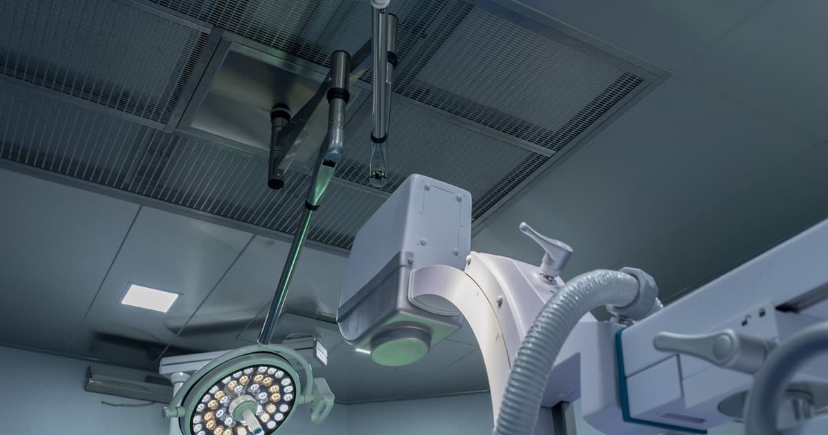
In a fictional case, a 42-year-old orthopedic surgeon operates on fractures with a C-arm several nights a week. In the corridor, the hemodynamics nurse mentions a colleague interventionalist treated for glioblastoma, and the radiology technician asks whether the thyroid shield is still worth it after so many years. The question that reaches the preceptor is not about cataract: it is about long-term cancer.
Fluoroscopy guides procedures in orthopedics, hemodynamics, interventional radiology, vascular surgery and bedside radiographs in the ICU. The team, physicians, nursing and technicians, stays close to the scatter source. This text gathers what large cohorts, case series and surveys show about brain and neck tumors, breast, thyroid and leukemia, makes the limits of each design clear and translates the evidence into practical protection. Cataract and staff skin are covered in the post on lens and skin in interventional radiology; ICRP 118 addresses tissue reactions, such as those of the lens (ICRP, 2012); here the focus is long-term neoplasia.
Key points
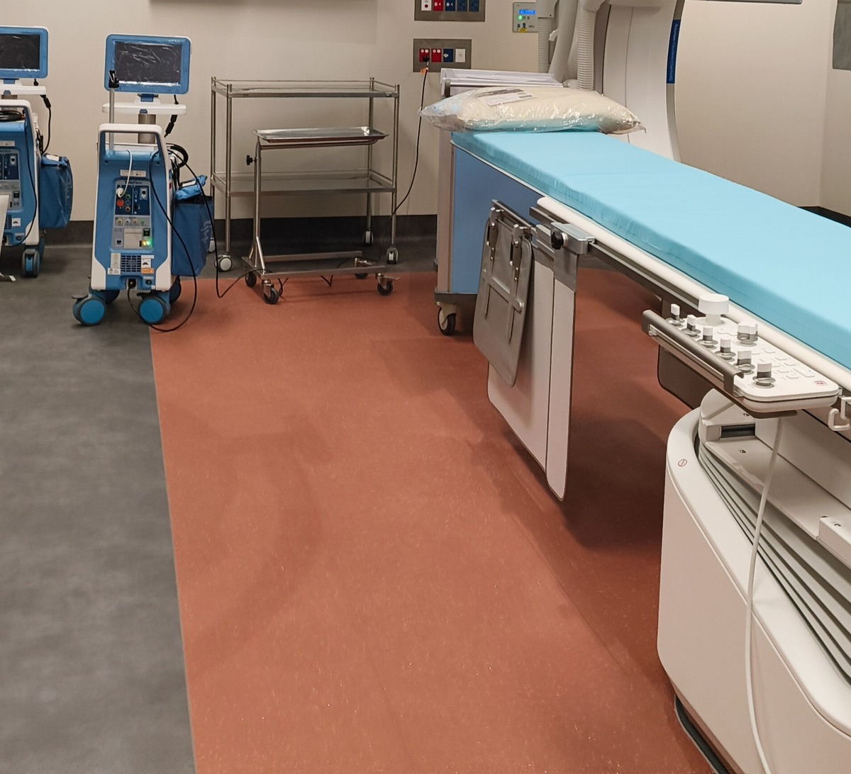
Fluoroscopy produces real-time images and spreads radiation through the room environment. Those who operate near the table, who hold the limb, who record the image and who assist in the ICU with a mobile unit receive scatter. The ALARA principle (as low as reasonably achievable) and the ICRP recommendations for procedures under guided fluoroscopy call for distance, shielding and reduced emission time (ICRP Publication 117).
The specialties with the longest fluoroscopy time include orthopedics (fractures, spine), hemodynamics and electrophysiology, interventional radiology, vascular surgery and ICU and emergency teams with portable radiography. Nursing and technicians spend years in the same room. Cumulative exposure over a career, and not a single day, is the object of the cohorts and surveys discussed below (our synthesis).
The gray (Gy) and the milligray (mGy) measure energy absorbed in tissue. The sievert (Sv) and the millisievert (mSv) express effective or equivalent exposure, useful for comparing scenarios. The INWORKS cohorts estimate risk per accumulated Gy; the USRT estimates occupational organ exposure in mGy. The authors' original units are kept in this text.
The best estimates of cancer risk after prolonged exposure to low amounts of radiation come from monitored nuclear workers, not from catheterization laboratories. They serve as a biological reference, with the caveat that the geometry of fluoroscopy (asymmetric scatter, partially unprotected head and trunk) differs from typical monitoring in the nuclear industry (our synthesis).
The INWORKS cohorts appear here as an explicit analogy: they are nuclear workers, not health care teams, and they serve to show the biological relationship between accumulated exposure and cancer. Richardson and colleagues updated the International Nuclear Workers Study (INWORKS) with 309,932 workers from France, the United Kingdom and the USA, 10.7 million person-years and 28,089 deaths from solid cancer (Richardson et al., 2023). The solid cancer mortality rate increased with accumulated exposure: excess relative risk of 0.52 per Gy (90 percent interval of 0.27 to 0.77), with a 10-year latency (Richardson et al., 2023). In the 0 to 100 mGy range, the association held and the interval widened. The authors describe the result as evidence of risk even at low accumulated exposures, typical of occupational settings.
In the previous analysis by the same collaboration, with 308,297 workers and 17,957 deaths from solid cancer, the excess relative risk was 0.47 per Gy (0.18 to 0.79) for solid cancer, with a mean colon exposure of 20.9 mGy among the exposed (Richardson et al., 2015). The cohort is not made up of health professionals; the extrapolation to the procedure room is biological, not directly empirical (our synthesis).
Leuraud and colleagues, using the same INWORKS base, analyzed mortality from leukemia and lymphoma. For leukemia excluding chronic lymphocytic leukemia, the excess relative risk was 2.96 per Gy (90 percent interval of 1.17 to 5.21), with a 2-year latency; for chronic myeloid leukemia, 10.45 per Gy (4.48 to 19.65) (Leuraud et al., 2015). The mean annual bone marrow exposure was low (about 1.1 mGy per year), and chronic lymphocytic leukemia showed no positive association (Leuraud et al., 2015). These numbers support the concern about prolonged occupational exposure, without proving the specific risk of hospital fluoroscopy (our synthesis).
The INWORKS cohorts measure workers with individual dosimetry over decades. The strength of the design is the large denominator and the measured exposure. The weakness, for the reader in the catheterization laboratory, is the geometry: the fluoroscopy beam is localized, the scatter is asymmetric, and the head, neck and hands may remain outside the apron. Therefore, INWORKS is an analogy, not a measurement of the room: they are nuclear workers. Using the INWORKS excess relative risk per Gy as the orthopedist's personal number would be a mistake. The correct use is biological: it confirms that prolonged exposure to low amounts of radiation is associated with solid cancer and leukemia; the magnitude in your room depends on emission time, protection and position (our synthesis).
The USRT brings the hospital closer, because the participants are radiology technologists. Even so, organ exposure was reconstructed from work history and models, not from a dosimeter on the lens or the breast at every shift. Wide intervals and self-reported FGIP call for a humble reading of the hazard ratios (Rajaraman et al., 2016).
The historical review by Linet and colleagues summarizes that the clearest excesses in medical workers occurred in those who started before 1950, especially leukemia, breast and skin, when protections were weaker (Linet et al., 2010).
The US Radiologic Technologists Study (USRT) follows technologists in the USA with questionnaires and cancer registries. Rajaraman and colleagues compared those who reported work with fluoroscopically guided interventional procedures (FGIP) between 1994 and 2008 with those who did not.
Brain cancer mortality was about twice as high among those who had FGIP (hazard ratio 2.55; 95 percent interval 1.48 to 4.40) (Rajaraman et al., 2016). The incidence of melanoma (1.30; 1.05 to 1.61) and of female breast cancer (1.16; 1.02 to 1.32) also rose; breast cancer mortality did not (1.07; 0.69 to 1.66) (Rajaraman et al., 2016). The incidence of all cancers combined, excluding non-melanoma skin, was at the limit (1.08; 1.00 to 1.17), and the authors warn about exposure estimated by self-report and about possible confounders (Rajaraman et al., 2016).
In a breast analysis in USRT, Preston, Kitahara and colleagues found a positive association between occupational radiation estimated in the breast and breast cancer risk, with an excess relative risk of 0.07 per 100 mGy (95 percent interval −0.005 to 0.19) (Preston et al., 2016). The association was clearer in women born before 1930, when the estimated mean annual exposure was much higher (about 37 mGy) than in later years (about 1.3 mGy) (Preston et al., 2016). The interval that nearly touches the null calls for caution: the signal exists, but precision is limited.
For thyroid, Kitahara and colleagues followed 89,897 technicians with 476 incident thyroid cancer diagnoses. The estimated mean thyroid exposure was 57 mGy. The excess relative risk per 100 mGy was −0.05 (95 percent interval from less than −0.10 to 0.34): the interval includes the null, and the result is inconclusive for excess thyroid cancer in adulthood with this exposure (Kitahara et al., 2018). This does not justify abandoning the thyroid shield: by the ALARA principle, it remains recommended even with inconclusive evidence, because it reduces organ exposure at low cost (our synthesis).
Pediatric patients and tomography appear in another text, about tomography in children and cancer risk. Follow-up after radiological accidents is in long-term follow-up after radiation exposure.
Roguin and colleagues gathered reports of brain and neck tumors in physicians who performed interventional procedures. In the expanded report, 31 physicians (23 interventional cardiologists, 2 electrophysiologists and 6 interventional radiologists) had worked long periods (mean latency about 23.5 years) (Roguin et al., 2013). Among the 26 physicians with a reported side, 22 tumors were on the left (85 percent), 3 on the right and 1 in the midline (Roguin et al., 2013). The earlier report already described a left predominance and recalled that the left side of the head is more exposed at the table (Roguin et al., 2012).
A case series does not estimate incidence or relative risk. There is no denominator of interventionalist-years, nor a formal comparison with the population. Reporting bias is expected: striking cases reach the authors; cases without tumor do not. Jackson discussed the same question and asked for caution before concluding causality from laterality alone (Jackson, 2012). The honest reading: the series generates a biologically plausible hypothesis and reinforces protection of the head and neck; it does not quantify the risk (our synthesis).
Chou and colleagues sent a questionnaire to women AAOS fellows. Among 505 responses (about 70 percent of those eligible), the standardized prevalence of any cancer was 1.85 times the expected (interval 1.19 to 2.76), and that of breast cancer, 2.9 times (1.66 to 4.71), a number that should be read as a hypothesis, not as an established risk (Chou et al., 2012). In the comparison between specialties, orthopedics kept the excess; urology and plastic surgery, with less weekly fluoroscopy, did not (Chou et al., 2015). Surgeons with cancer may have responded more to the questionnaire (response bias), and the comparison with the general population also distorts; the design does not prove that fluoroscopy is the cause, but it justifies adequate breast shielding (our synthesis).
Valone and colleagues used an anthropomorphic phantom and showed that the upper outer quadrant of the breast can be poorly covered depending on the size and type of apron, the surgeon's position and the orientation of the C-arm (Valone et al., 2016). Hayda and colleagues reviewed occupational risks for orthopedists and reiterate the ALARA principle, standardized education and dosimetry (Hayda et al., 2018).
In an Italian orthopedic hospital with protection described as poor, Mastrangelo and colleagues found a cumulative cancer incidence of 29 percent (9 of 31) among orthopedists, versus 4 percent (7 of 158) in the unexposed; the regression linked work as an orthopedist to tumor risk (Mastrangelo et al., 2005). The sample is small and the context of inadequate protection limits generalization.
Andreassi and colleagues compared 466 professionals exposed in hemodynamics and electrophysiology with 280 unexposed. In highly exposed physicians, the adjusted odds ratio for cancer was 4.5 (interval 0.9 to 25; p = 0.06): the interval includes the null and the result is inconclusive for cancer, although other conditions were associated with exposure (Andreassi et al., 2016).
Orme and colleagues, in a multicenter Mayo Clinic survey with 1,543 responses, found no difference in cancer prevalence between those who took part in procedures involving radiation and those who did not (9 percent in both groups) (Orme et al., 2015). Musculoskeletal pain was greater in the exposed. Read together, Andreassi suggests a signal that does not reach significance and Orme finds no difference; neither closes the question, and the balance between them is the honest picture of the evidence in hemodynamics (our synthesis). Klein and SCAI described, in a survey of 314 interventionalists, workload limitation due to exposure and concerns about cataract and cancer, without estimating population relative risk (Klein et al., 2015).
The table below summarizes the design and what each type of study allows one to conclude (our synthesis).
| Type of evidence | Examples | What it measures | Main limitation |
|---|---|---|---|
| Cohort with individual dosimetry | INWORKS (Richardson, Leuraud) | Cancer mortality vs cumulative exposure in Gy | Few are health professionals; geometry differs from fluoroscopy |
| Technologist cohort (USRT) | Rajaraman; Preston; Kitahara | Incidence and mortality vs estimated FGIP or mGy | Self-reported procedures; wide intervals |
| Case series | Roguin 2012 and 2013 | Description and laterality of tumors | No denominator; reporting bias; risk estimate unavailable |
| Prevalence survey | Chou; Andreassi; Orme; Klein | Self-reported prevalence vs controls or population | Response bias; causality not established |
| Phantom / bench dosimetry | Valone | Relative exposure of trunk regions | Measures only relative exposure, with no clinical cancer outcome |
Sources: Richardson et al., 2023; Leuraud et al., 2015; Rajaraman et al., 2016; Roguin et al., 2013; Chou et al., 2012; Valone et al., 2016.
In the ICU and the emergency department, the mobile X-ray machine and the portable C-arm place nursing staff and technologists a few centimeters from the patient. Whoever holds the cassette, whoever stabilizes the head and whoever stays at the head of the bed during emission receives scatter. The culture of "just one quick shot" without an apron is the opposite of ALARA. Announcing the emission, moving away those who are not essential and wearing protection even on crowded shifts are team measures, not those of an individual hero (our synthesis).
Orme and colleagues note that musculoskeletal pain was greater in technologists and nurses than in attending physicians in the interventional laboratory, which shows that the physical load of the apron and of long postures is also occupational (Orme et al., 2015). Cancer and spine do not compete: both call for safe work design.
Three reading rules avoid alarmism and complacency (our synthesis).
First: a case series generates a hypothesis, not incidence. Roguin documents tumors and laterality; it does not say how many interventionalists per thousand will have a tumor.
Second: reporting and response bias. Those who had cancer respond more to surveys about cancer; those who are healthy disappear. Comparing the prevalence of female surgeons with the general population mixes reproductive factors, screening and occupation.
Third: a confidence interval that includes the null is inconclusive. Thyroid in the USRT (−0.05 per 100 mGy, interval crossing zero) (Kitahara et al., 2018) and cancer in the Andreassi study (odds ratio 4.5; 0.9 to 25) (Andreassi et al., 2016) prove neither the absence nor the presence of risk. Absence of evidence is not evidence of absence.
A useful conversation in the department separates three layers. Layer 1: the biology (ionizing radiation can initiate cancer; occupational cohorts show an association). Layer 2: the epidemiology of your specialty (case series and surveys generate hypotheses; technician cohorts suggest signals in brain, breast and melanoma; thyroid remained inconclusive). Layer 3: the action (time, distance, shielding, dosimetry, pregnancy). Mixing the three layers into a single "we are going to get cancer" or a single "there is no proof, so ignore it" undermines safety (our synthesis).
Reading evidence with study design and interval in mind is daily training: whoever reads Roguin must avoid turning a case series into an incidence rate, and whoever reads Chou must remember who answered the questionnaire (our synthesis).
The editorial synthesis of this post is a working hypothesis: prolonged occupational exposure to fluoroscopy is biologically capable of raising the risk of some cancers; the magnitude in modern procedure rooms, with correct protection, remains poorly quantified; the practical answer is to reduce exposure without abandoning necessary procedures.
ICRP 117 organizes protection in procedures under guided fluoroscopy: justify the use, optimize the emission and shield the team (ICRP Publication 117). In daily practice:
Before the first case of the day: check the apron (size and type), thyroid shield, glasses if indicated, screen in position and dosimeter in the right place. During the case: pulsed fluoroscopy, hands out of the primary beam, distance when the image allows, fewer cine runs "out of habit". After the case: store the dosimeter, report high values and log equipment failures. In teams with a high load of spine and trauma, education on breast and thyroid shielding should enter residency with the same seriousness as surgical technique (Hayda et al., 2018; Valone et al., 2016).
Vascular surgery and hemodynamics share the same scatter physics. The difference lies in the annual volume of procedures and in the chronic proximity to the table. Those who accumulate thousands of procedures over two decades are the target audience of the Roguin series and the SCAI surveys, even though these designs do not measure incidence (Roguin et al., 2013; Klein et al., 2015).
ICU and bedside radiography: whoever holds the cassette or stays at the head of the bed receives scatter. Keeping away those who are not essential, wearing an apron and announcing the emission before the shot are cheap and effective gestures (our synthesis).
Team well-being is not limited to radiation. Fatigue, workload and safety culture appear in texts such as working sick: physician presenteeism and moral injury. The patient's clinical oncology, distinct from occupational risk, is in posts such as breast cancer: treatment sequence.
In NEXO, the case of the day is fictional, with a real clinical presentation and up to six progressive clues, and you propose the diagnosis by looking for the ICD-10 code. Reading an occupational study calls for the same care with design, bias and confidence interval that the game calls for when putting clues together without closing too early.
The limits on using the game are described on the Economy page of NEXO. If you research the teaching of clinical reasoning, the page for Researcher explains how to get in touch. Professionals who write or review cases find information on Curator and on Creators, and institutions can learn about the proposal on the partnerships page.
The series describes tumors and left-sided laterality that is biologically plausible, with no population denominator and no relative risk (Roguin et al., 2013; Jackson, 2012). It does not prove the individual risk of each interventionalist.
In the USRT, mortality from brain cancer and the incidence of melanoma and breast cancer rose among those who reported FGIP; other sites did not (Rajaraman et al., 2016). Exposure was estimated from work history.
Yes. The thyroid analysis was inconclusive (an interval that includes the null), not proof that there is no risk (Kitahara et al., 2018). Under the ALARA principle, the shield remains recommended even with inconclusive evidence: it is cheap and reduces exposure of the organ (Hayda et al., 2018).
With advance declaration of pregnancy, reinforced shielding, dosimetry and, if needed, reassignment. Details of fetal counseling are in the post on radiation in pregnancy.
Only by reference. Thresholds, glasses and staff skin are in lens and skin in interventional radiology.
Play the case of the day in NEXO and train your reasoning with progressive clues.