



Armed conflict and humanitarian crisesRadiation exposure
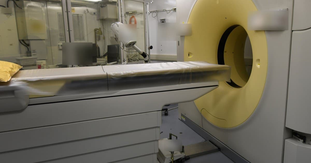
In a fictional case, a 4-year-old girl arrives at the emergency department at 11 p.m. with abdominal pain for one day, vomiting and low-grade fever. The examination is doubtful, the mother is distraught and the resident considers ordering an abdominal CT scan to "not miss anything". The supervising physician asks first: what will the CT scan answer that ultrasound and a repeat assessment in a few hours will not answer? And, if it does answer, what changes in management tonight?
The question does not come from fear of radiation, but from clinical reasoning. Over the past decade, cohorts with millions of children have measured, with estimates of organ exposure in mGy, what happens after CT scans in childhood. This text gathers what they show about leukemia and brain tumors, where the limits of these studies lie and why the clinical question, and not habit, should decide the examination.
Key points
Computed tomography (CT) is one of the most useful diagnostic tools in medicine, but it exposes the patient to more ionizing radiation than most imaging examinations. The authors of the Dutch cohort summarize the concern with three arguments: a child may receive more radiation than needed if the protocol is an adult one, is more sensitive to the carcinogenic effects of radiation and has more years of life ahead for a late effect to appear (Meulepas et al., 2019).
The same authors recall that leukemia and brain tumors are the most common radiation-induced neoplasms in children and young adults. The thyroid is also very sensitive, but thyroid cancer is too rare in this age range for an excess to appear in short follow-up cohorts. For this reason, most cohorts of CT scans in childhood looked first at leukemia, measured by radiation in the bone marrow, and at brain tumor, measured by radiation in the brain.
The mGy (milligray) measures the energy absorbed by an organ, such as the marrow or the brain, and is the unit that recent cohorts estimate for each examination. The mSv (millisievert) is the so-called effective exposure, a single number for the whole body, more useful for comparing examinations than for calculating the risk to one organ. In the Australian cohort, each CT scan corresponded on average to 4.5 mSv of effective exposure (Mathews et al., 2013).
Risk is usually described as excess relative risk (ERR) per 100 mGy, that is, how much the risk increases, relative to baseline, for each 100 mGy accumulated in the organ. An ERR of 1 per 100 mGy means that 100 mGy would double the baseline risk. Because childhood cancers are rare, doubling a very low risk still results in a low absolute risk, and this distinction runs through the whole text.
The study by Pearce and colleagues is described by the authors as a retrospective cohort of patients without previous cancer who had their first CT at public health system services in England, Wales and Scotland between 1985 and 2002, before age 22 (Pearce et al., 2012). To avoid counting CT scans done because of the cancer itself, leukemia follow-up began 2 years after the first CT.
Among 178,604 patients, 74 had leukemia. The excess relative risk was 0.036 per mGy of radiation to the bone marrow, with a 95 percent confidence interval of 0.005 to 0.120 (Pearce et al., 2012). Compared with those who received less than 5 mGy, those who accumulated 30 mGy or more had a relative risk of leukemia of 3.18 (1.46 to 6.94) (Pearce et al., 2012). The authors translate the finding as follows: two or three head CTs, with about 50 mGy accumulated in the marrow, could nearly triple the risk of leukemia.
EPI-CT combined and expanded national cohorts to overcome the limits of smaller studies. The authors describe a multinational cohort of 948,174 people who had CT scans before age 22 in nine European countries, with active bone marrow radiation estimated from the region examined, the patient's characteristics, the period and the inferred technical parameters (Bosch de Basea et al., 2023).
With 790 hematological malignancies, the excess relative risk was 1.96 per 100 mGy, with an interval of 1.10 to 3.12 (Bosch de Basea et al., 2023). The estimates were similar for lymphoid and myeloid malignancies. In the authors' translation, for every 10,000 children examined today, with an average of 8 mGy to the marrow, 1 to 2 should develop a hematological malignancy attributable to radiation in the following 12 years.
Smith-Bindman and colleagues describe a retrospective cohort of 3,724,623 children born between 1996 and 2016 in six United States health systems and in Ontario, Canada, followed up to age 21 (Smith-Bindman et al., 2025). The difference from the European cohorts is that the study included all imaging modalities and compared children exposed and not exposed since birth.
A total of 2,961 hematological malignancies were diagnosed. Accumulated radiation to the marrow was associated with risk, with an excess relative risk of 2.54 per 100 mGy (1.70 to 3.51) (Smith-Bindman et al., 2025). In a modeling calculation, the authors estimated that 10.1 percent (5.8 to 14.2) of the cohort's hematological malignancies could be attributed to radiation from imaging exams, with a greater weight for the modalities with the highest exposure, such as CT (Smith-Bindman et al., 2025). The average among children with at least 1 mGy was 14 mGy, which the authors themselves compare to a head CT.
The result is not identical in all cohorts. In the Dutch cohort, accumulated radiation to the marrow averaged 9.5 mGy, and the risk of leukemia was not associated with it, with 44 diagnoses (Meulepas et al., 2019). With few events and low average exposure, a study of this size has little ability to detect a small effect, and the absence of association there should be read as an inconclusive result, not as proof that there is no risk (our synthesis).
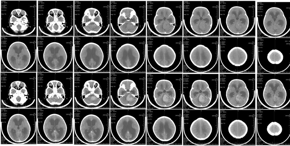
In the British cohort, follow-up for brain tumor began only 5 years after the first CT, precisely to rule out exams prompted by a tumor not yet diagnosed. Among 176,587 patients, 135 had a brain tumor, with an excess relative risk of 0.023 per mGy (0.010 to 0.049) (Pearce et al., 2012). Compared with less than 5 mGy, those who received 50 to 74 mGy to the brain had a relative risk of 2.82 (1.33 to 6.03) (Pearce et al., 2012).
The Dutch cohort is described by the authors as a national retrospective cohort of 168,394 children who had at least one CT before age 18, between 1979 and 2012 (Meulepas et al., 2019). Accumulated radiation to the brain averaged 38.5 mGy and was associated with the risk of malignant and benign brain tumors together, with an excess relative risk of 0.86 per 100 mGy (0.20 to 2.22), in 84 diagnoses (Meulepas et al., 2019).
The Australian study by Mathews and colleagues is described as a population-based data linkage study, with 10.9 million people identified in public health system records (Mathews et al., 2013). Among 680,211 people exposed to CT at least one year before any cancer, the overall cancer incidence was higher than among the unexposed, with an incidence rate ratio of 1.24 (1.20 to 1.29) (Mathews et al., 2013). The ratio rose by 0.16 with each additional CT, and the effect was greater the younger the child at the first scan (Mathews et al., 2013). The study counted scans and did not estimate the radiation received by each organ.
A later analysis of the same Australian records estimated the radiation dose to the brain. Among 611,544 young people exposed before age 20, with a mean of 44 mGy to the brain, the excess relative risk of brain cancer was 0.8 per 100 mGy (0.57 to 1.06) (Smoll et al., 2023).
In Korea, Han and colleagues describe a national cohort based on health insurance data, with 1,380,896 people under 20 who had a CT between 2007 and 2015, excluding those with congenital anomalies or prior cancer (Han et al., 2025). Risk was expressed per standard deviation of organ radiation dose, not per 100 mGy. For brain cancer, the hazard ratio was 1.201 (1.177 to 1.225), and for leukemia, 1.074 (1.053 to 1.100) (Han et al., 2025). Because it uses another unit, this number cannot be added to or directly compared with the ERR per 100 mGy of the other cohorts (our synthesis).
A recent systematic review gathered seven large cohorts from Europe, Asia and Australia, with more than 11 million people. Among the studies with an estimate of organ radiation dose, the excess relative risk per mGy ranged from 0.033 to 0.045 for leukemia and from 0.006 to 0.023 for brain tumor (Almatova et al., 2026). Risk was higher in those exposed younger, especially under age 5. The reviewers warn that counting scans is not the same as measuring organ radiation dose, and that indication bias was identified, although adjusted and latency analyses generally kept the association.
CT cohorts have two main methodological enemies, and every author read here devotes space to them.
Reverse causality happens when the cancer prompts the scan, not the other way around. A brain tumor can cause headache or seizures years before diagnosis, and the child has the CT because of the tumor still hidden. The Australian authors acknowledge this explicitly: the brain cancer may have caused the scan. To measure the problem, they repeated the analyses excluding brain cancers that came after a head CT, and the excess of other cancers persisted (Mathews et al., 2013).
The main defenses are latency periods and exclusions. Pearce began counting leukemias 2 years and brain tumors 5 years after the first CT (Pearce et al., 2012). In North America, with latencies of 6 and 24 months, the raised risk persisted, and the authors argue that these malignancies are usually diagnosed quickly (Smith-Bindman et al., 2025). In EPI-CT, the authors consider reverse causality unlikely because the estimates remained raised with longer latencies (Bosch de Basea et al., 2023).
Even so, the doubt does not disappear completely. Pearce and colleagues admit that they cannot rule out this bias and that the increase in brain tumor risk greater than expected from atomic bomb survivors may indicate some residual bias, despite the long exclusion period (Pearce et al., 2012). In Australia, the analysis with brain radiation dose estimated that 40 percent (29 to 50) of brain tumors arising more than 2 years after a CT would be attributable to radiation, and not to reverse causality (Smoll et al., 2023). In this estimate, the remainder of these tumors would not be explained by CT radiation (our synthesis).
The second problem is predisposition. Children with syndromes that increase cancer risk, such as tuberous sclerosis, undergo more head imaging because of the syndrome itself. In the Dutch cohort, the authors treated tuberous sclerosis as a potential confounder, and excluding these patients did not substantially change the risk (Meulepas et al., 2019). EPI-CT excluded malignancies recorded as linked to therapy or to predisposing syndromes (Bosch de Basea et al., 2023).
Even so, the incidence of brain tumor in the Dutch cohort was higher than in the general population, and the authors call for cautious interpretation (Meulepas et al., 2019). In Korea, the authors point out that the retrospective design and administrative data can bring selection bias and unmeasured confounders (Han et al., 2025). People who are scanned many times almost always have an underlying disease, and separating the effect of radiation from the effect of the disease is the hardest point in these studies (our synthesis).
A British study extracted the medical conditions from 37 807 reports of children and young people without cancer. Hydrocephalus appeared in 19 percent of all CT scans and in 59 percent of those repeated 5 times or more in the same year (Journy et al., 2016). Suspected or diagnosed tumor appeared in only 5 percent of CT scans (Journy et al., 2016). The authors conclude that children with a ventricular shunt may accumulate a lot of radiation early in life, precisely at the age of greatest sensitivity.
The number that matters for the family and for the clinical decision is the absolute risk. In the British cohort, in children under 10 years old, an estimated one extra case of leukemia and one of brain tumor occur per 10 000 head CT scans in the 10 years following the first exam (Pearce et al., 2012). The authors state clearly that the clinical benefits must outweigh this small risk, but that radiation should be kept as low as possible.
In Australia, the absolute excess for all cancers combined was 9.38 per 100 000 person-years (Mathews et al., 2013). In the brain cancer analysis, about 6391 people would need to be exposed to cause one extra brain cancer, and only 3.7 percent of all brain cancers in the country would be attributable to CT (Smoll et al., 2023). The authors note that this fraction would be higher in countries that perform more CT scans in children.
A British projection estimated a lifetime excess risk of 0.3 to 1 per 1000 head CT scans and of 1 to 5 per 1000 CT scans of other regions, with practice from 2000 to 2008 (Journy et al., 2017). This is a modeling study, not a cohort: the number comes from risk models applied to exposure estimates. The authors conclude that the risk of a single exam is low compared with the baseline risk, but that small increases in the annual number of CT scans would raise the total of future cancers considerably.
For the family, it helps to separate three ideas (our synthesis). The first is that the risk exists, appears in cohorts from several countries and grows with accumulated exposure. The second is that, for a well-indicated exam, it is small next to the risk of leaving a serious disease undiagnosed. The third is that the team can reduce it by requesting only the exam that changes management and preferring methods without radiation when they answer the same question. To check whether the family understood, asking them to repeat it in their own words works better than asking whether it was clear.
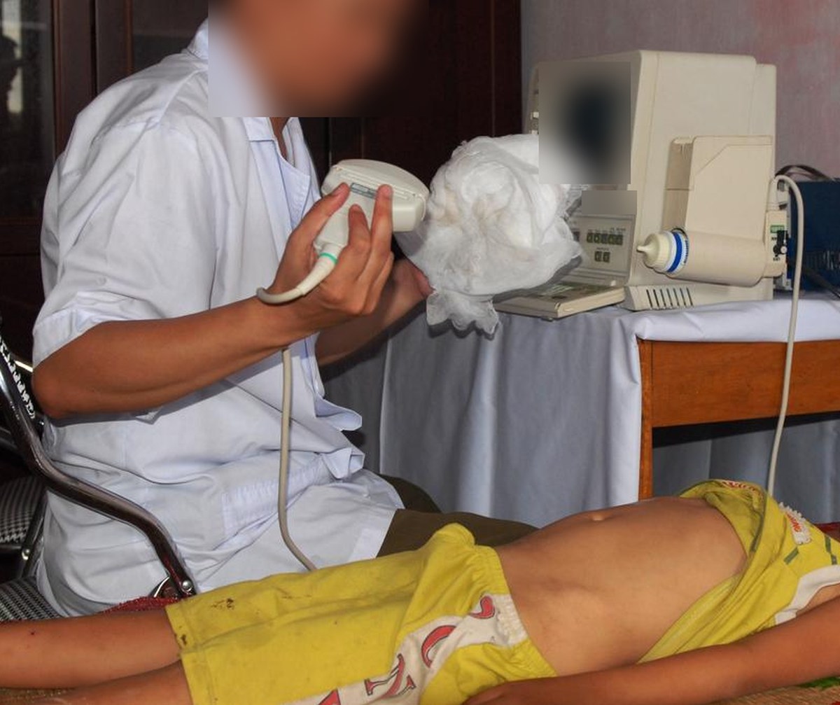
The authors of the Korean editorial summarize the principle: the goal is not to restrict appropriate imaging, but to eliminate exams without justification, which expose the patient to risk without diagnostic benefit. Imaging should be done only when it is expected to change management (Hwang et al., 2025). The same text cites an audit in seven European countries in which almost a quarter of CT requests did not follow evidence-based guidelines.
Four questions help organize the decision (our synthesis). Which hypothesis do I need to confirm or rule out? Will the result change what I do today: how to manage, admit, discharge or change treatment? Is there an exam without ionizing radiation, such as ultrasound or MRI, that answers the same question safely? And would a clinical reassessment in a few hours resolve the doubt without any exam at all?
In the case of the onset, these questions do not forbid CT. If the ultrasound is not conclusive and the child worsens, tomography may be the right exam, and the radiation from a well-indicated exam is no reason to delay a serious diagnosis. The point is that the exam comes after the question, not in its place.
Mild head trauma is the situation where unnecessary CT in children is most discussed. The Korean editorial reports, citing earlier studies, that 25 to 30 percent of CT scans for mild head trauma in pediatric emergency departments would be unnecessary, despite clinical rules to identify who is at low risk (Hwang et al., 2025). The decision rules and who needs tomography are in the text on mild head injury and who needs tomography, and are not repeated here.
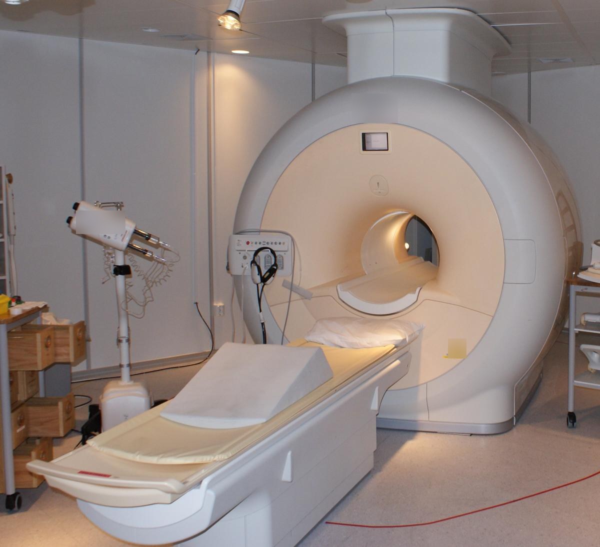
For neurological complaints without trauma, a pediatric hospital in the United States routinely offered four rapid MRI protocols in the emergency department. The authors describe a retrospective study comparing two 9-month periods, before and after the change. Use of head CT fell 21.5 percent (17.5 to 25.5) (Ramgopal et al., 2020). No rapid MRI with follow-up by full MRI failed to show a lesion.
The switch had a disadvantage: time to imaging and length of stay in the emergency department were longer with rapid MRI, and 3.6 percent of these exams failed, almost all due to the child moving (Ramgopal et al., 2020). It is a single-center study that compared periods before and after, without randomization, and the result depends on having MRI available at night.
Awareness campaigns help, but the effect is hard to measure. A study using health insurance data from children with kidney stones, with a difference-in-differences design, saw CT use fall after the start of the Image Gently campaign, but also fall among adults, the control group (Streur et al., 2019). The authors conclude that the decline cannot be attributed mainly to the campaign.
If CT is the right exam, exposure can still be lower. A British survey of 1073 sets of films from 36 hospitals showed that the estimated radiation to the brain in a head CT fell from 62 mGy before 1990 to about 30 mGy after 2000 (Lee et al., 2016). In chest CT for children aged 0 to 4 years, radiation to the lung fell from 28 to 4 mGy, with parameters adjusted for age (Lee et al., 2016). In the British projection, risk per exam fell 50 to 70 percent between 1990 and 2008 because of this reduction (Journy et al., 2017).
In practice, this means requesting the exam while stating the clinical question, so that radiology chooses the region and the protocol, and avoiding repeating a CT that another service has already done (our synthesis). In children who will need many exams over their lifetime, such as those with a ventricular shunt, the text on long-term follow-up after radiation exposure covers monitoring, and for pregnant women the text on radiation in pregnancy and counseling applies.
| Study | Design | Population | Main finding |
|---|---|---|---|
| Pearce, 2012 | Retrospective cohort | 178,604 young people in Great Britain | Leukemia and brain tumor increase with accumulated mGy (Pearce et al., 2012) |
| Mathews, 2013 | Data linkage, population-based | 10.9 million in Australia (Mathews et al., 2013) | More cancer in the exposed, greater with more exams |
| Meulepas, 2019 | National retrospective cohort | 168,394 children in the Netherlands | Brain tumor associated; leukemia without association (Meulepas et al., 2019) |
| Bosch de Basea, 2023 | EPI-CT multinational cohort | 948,174 in nine countries | Hematologic malignancies increase with mGy to the marrow (Bosch de Basea et al., 2023) |
| Smith-Bindman, 2025 | Retrospective cohort | 3,724,623 children in the US and Ontario | Hematologic malignancies increase with imaging radiation (Smith-Bindman et al., 2025) |
| Han, 2025 | National cohort | 1,380,896 in Korea | Association per standard deviation of organ exposure (Han et al., 2025) |
Sources: the risks come from estimates of organ radiation in mGy, except in Mathews, which counted exams, and in Han, which used standard deviation. Data from Pearce et al., 2012, Mathews et al., 2013, Meulepas et al., 2019, Bosch de Basea et al., 2023, Smith-Bindman et al., 2025 and Han et al., 2025.
In NEXO, the case of the day is fictional, with a real clinical presentation and up to six progressive clues, and you propose the diagnosis by looking for the ICD-10 code. Deciding whether an exam changes management is the same exercise of putting together signs, symptoms and history that appears in the text on Signs, symptoms and history: how to interpret what the patient brings.
The limits of using the game are described on the NEXO Economy page. If you research clinical reasoning teaching, the Researcher page explains how to get in touch. Professionals who write or review cases find information on Curator and Creators, and institutions can learn about the proposal on the partnerships page.
The risk from a single exam is small. In the British cohort, an estimated one extra case of leukemia and one of brain tumor per 10,000 head CTs over ten years is estimated, in children under 10 (Pearce et al., 2012). The studies show an association in the population, and do not allow saying which child will develop cancer because of an exam.
In part, and the authors know this. Latencies, exclusions and sensitivity analyses reduce reverse causality and the effect of predisposing syndromes, but do not eliminate them completely (Meulepas et al., 2019).
Radiography exposes to far less radiation. In the North American cohort, the modalities with the highest exposure, such as CT, weighed more in the fraction of attributable malignancies (Smith-Bindman et al., 2025).
The studies here looked at medical exams. Occupational and environmental exposures appear in other texts, such as those on interventional radiology and the team, cosmic radiation in crews and potassium iodide by age.
Play the case of the day on NEXO and train your reasoning with progressive clues.