NephrologyEmergency medicineClerkships and residency
Metabolic acidosis: anion gap step by step



Table of contents
- Why read blood gas step by step
- The first steps: pH, primary disorder and compensation
- Step 4: the anion gap and its pitfalls
- Step 5: the delta and the hidden disorder
- High anion gap: the causes
- Normal anion gap: bicarbonate loss and the kidney
- Treat the cause, and when to think of bicarbonate
- How NEXO helps train this reasoning
- Frequently asked questions
- Take-home messages
- References
In a fictional case, a 58-year-old woman arrives at the emergency department with vomiting, weakness and rapid breathing. She has diabetes and takes an SGLT-2 inhibitor. Her blood glucose is not very high, and so nobody thought of ketoacidosis. Venous blood gas shows low pH and bicarbonate of 12 mmol/L. The intern looks at the numbers and does not know where to start.
Blood gas is intimidating because it brings many numbers at once. Read in order, it becomes a sequence of clues, each one narrowing the hypotheses. A nephrology review in intensive care summarizes the reading in five steps and says that, done systematically, it becomes simple (Achanti and Szerlip, 2023).
This text goes through those steps as an exercise in reasoning by clues: the primary disorder, the expected compensation, the anion gap corrected for albumin, the delta and the causes of each pattern. At the end, it returns to the case and shows what each step revealed.
Key points
- The reading starts with the history and the pH, continues with compensation and the anion gap and ends with the delta, which reveals a second hidden disorder (Achanti and Szerlip, 2023).
- In metabolic acidosis, the Winters formula estimates the expected PCO2 as 1.5 times the bicarbonate plus 8, with a margin of 2 (Duška et al., 2026).
- The normal value of the anion gap depends on the laboratory method, and low albumin hides unmeasured anions (Lee et al., 2006; Kraut and Nagami, 2013).
- The anion gap has low sensitivity for mildly raised lactate, and so lactate should be measured (Glasmacher and Stones, 2017).
- Diarrhea, bicarbonate loss through the kidney and large volumes of saline cause acidosis with a normal anion gap (Achanti and Szerlip, 2023; Hamm et al., 2015).
- Sodium bicarbonate in severe acute acidosis reduced the need for renal replacement therapy in patients with acute kidney injury, with no clear effect on 28-day mortality (Lind et al., 2026).
Summary
- Why read blood gas step by step
- The first steps: pH, primary disorder and compensation
- Step 4: the anion gap and its pitfalls
- Step 5: the delta and the hidden disorder
- High anion gap: the causes
- Normal anion gap: bicarbonate loss and the kidney
- Treating the cause, and when to think of bicarbonate
- How NEXO helps train this reasoning
- Frequently asked questions
- Take-home messages
Why read blood gas step by step
A problem on any shift
Metabolic acidosis appears in very different causes, from diarrhea in a child to sepsis in a hospitalized adult. The World Health Organization (WHO) estimates that diarrhea kills about 443 thousand children under 5 years old per year, and that it leaves the body without the water and salts needed to survive (WHO, diarrheal disease). The same organization records 830 million people with diabetes in 2022 (WHO, diabetes), and ketoacidosis is one of the acute complications of this disease.
In intensive care, 8 to 14 percent of patients arrive with moderate metabolic acidosis, with pH below 7.30 (Lind et al., 2026). Knowing how to read blood gas, therefore, is a basic skill for any doctor, in any country.
The kidney and the lung share the work
The body produces acid every day. The kidneys regenerate the bicarbonate consumed by excreting acid, about 70 mEq per day under usual conditions, in the form of titratable acidity and ammonium (Hamm et al., 2015). When the acid load or the loss of base is too great, as in severe diarrhea, the kidneys cannot keep up and metabolic acidosis sets in (Hamm et al., 2015).
The lung responds faster. By eliminating more CO2, it reduces PCO2 and limits the fall in pH. This response has a predictable size, and it is precisely this prediction that allows mixed disorders to be discovered.
Blood gas as a game of clues
Each step of the reading works as a new clue. The first narrows the problem to an acidosis or an alkalosis; the second says whether it is metabolic or respiratory; the third shows whether there is a second disorder, and so on. This way of thinking is the same one we use to train differential diagnosis with clues: each piece of information changes the probability of the hypotheses, as in reading tests by pre-test probability.
The first steps: pH, primary disorder and compensation
Step 1: history and physical examination
Reading starts before the blood gas. History and physical examination give the clues to an acid-base disorder and guide the test order (Achanti and Szerlip, 2023). Vomiting, diarrhea, diabetes, medication use, kidney failure, shock and intoxications already suggest which disorders to look for.
A recent algorithm describes this step as estimating the pretest probability and severity (Duška et al., 2026). Extreme pH values, below 7.2 or above 7.6, call for rapid intervention (Duška et al., 2026).
Step 2: pH and the primary disorder
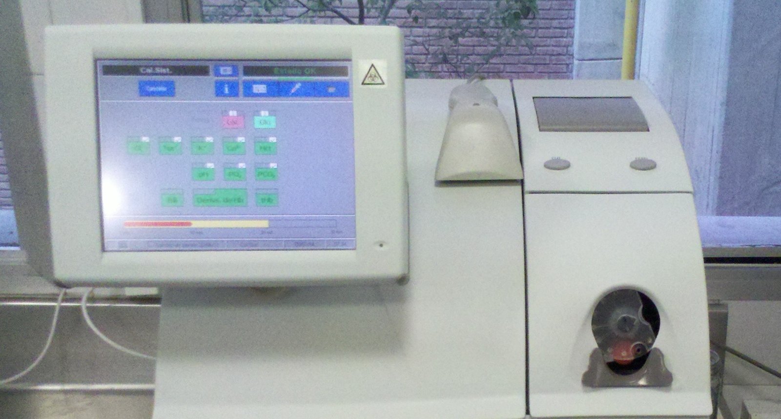

The blood gas shows whether there is acidemia or alkalemia and whether the disorder is metabolic or respiratory. In the stable patient, the difference between arterial and venous samples is small, and both work; in shock, the review recommends the arterial sample (Achanti and Szerlip, 2023).
A quick rule helps. If PCO2 and bicarbonate move in the same direction, there is usually a single disorder; if they move in opposite directions, there is a mixed disorder or a laboratory error (Achanti and Szerlip, 2023).
Step 3: the expected compensation
In metabolic acidosis, the Winters formula estimates the expected PCO2 as 1.5 times the bicarbonate plus 8, with a margin of 2 mmHg. A PCO2 above the predicted value indicates insufficient compensation or associated respiratory acidosis; a PCO2 below it indicates associated respiratory alkalosis (Duška et al., 2026).
This step has direct clinical value. When compensation fails, it may signal respiratory muscle fatigue and an imminent need for intubation (Duška et al., 2026). Compensation also almost never brings pH back to normal; a normal pH with low bicarbonate suggests another disorder (Achanti and Szerlip, 2023).
Step 4: the anion gap and its pitfalls
The calculation
The anion gap should be calculated whatever the pH, as sodium minus the sum of chloride and total CO2 (Achanti and Szerlip, 2023). The review notes that total CO2 comes from the biochemistry panel, includes bicarbonate, carbonate and dissolved CO2, and is usually 2 mEq higher than the bicarbonate calculated in the blood gas (Achanti and Szerlip, 2023).
The "gap" exists because there are more unmeasured anions, especially albumin, than unmeasured cations (Achanti and Szerlip, 2023). When an organic acid such as lactate or beta-hydroxybutyrate accumulates, its anion enters that space and the gap rises.
The normal value depends on the laboratory
The classic value of 12 ± 4 mEq/L came from the era when sodium was measured by flame photometry. With ion-selective electrodes, used since the 1980s, the normal value fell to about 6 ± 3 mEq/L (Lee et al., 2006). A review that compared textbooks and electronic sources found different normal ranges among them (Kraut and Nagami, 2013).
In practice, the intensive care review proposes considering a gap between 6 and 12 mEq/L normal and using 10 as a reference (Achanti and Szerlip, 2023). The best approach is to know the method and range of your own laboratory and, when possible, the patient's baseline gap (Kraut and Nagami, 2013).
Correction for albumin
Albumin is the main unmeasured anion, and hypoalbuminemia lowers the gap. For each 1 g/dL drop in albumin, the review advises adding 2.5 to the anion gap (Achanti and Szerlip, 2023). Without this correction, the malnourished, cirrhotic or septic patient may have an organic acid hidden under an apparently normal gap (Kraut and Nagami, 2013).
An anion gap above 18, whatever the pH, almost always indicates an organic acid (Achanti and Szerlip, 2023). The higher the gap, the greater the chance of identifying the cause.
The limit of lactate
The anion gap often fails to show mild lactic acidosis. In a group of 272 emergency patients, only 58 percent of those with lactate above 2.5 mmol/L had a gap outside the normal range (Kraut and Nagami, 2013). A meta-analysis concluded that the gap has low sensitivity for lactate above 2.5 mmol/L, but good discrimination for lactate above 4 mmol/L, with an area under the curve of 0.87 (Glasmacher and Stones, 2017).
The consequence is simple: lactate must be measured directly, even with a corrected gap (Achanti and Szerlip, 2023). In sepsis screening, this measurement is part of the first hour of care.
A low gap is also a clue
A low gap often goes unnoticed, but it can be the first or the only sign of a disease such as paraproteinemia. Some paraproteins, such as the IgG of multiple myeloma, have a positive charge and increase the unmeasured cations (Lee et al., 2006).
Step 5: the delta and the hidden disorder
The logic of the delta
If the organic acid is buffered only by bicarbonate, each 1 mEq/L rise in the gap should correspond to a 1 mEq/L fall in total CO2. To test this, the rise in the gap above normal is added to total CO2, and the result estimates the bicarbonate the patient would have without the acid (Achanti and Szerlip, 2023).
- If this estimated bicarbonate is above 28 mEq/L, there is probably an associated metabolic alkalosis (Achanti and Szerlip, 2023).
- A gap above 30, with a delta above 20, almost always indicates underlying metabolic alkalosis (Achanti and Szerlip, 2023).
- If the estimated bicarbonate is still low, there may be a normal gap acidosis added to the high gap acidosis.
Why the ratio varies
The relationship between the rise in the gap and the fall in bicarbonate changes with the acid. In human studies, it was 1 to 1 in ketoacidosis and, on average, 1.5 in lactic acidosis (Kraut and Nagami, 2013). Part of the filtered anions is excreted in the urine, which reduces the gap without correcting the bicarbonate (Kraut and Nagami, 2013).
That is why the delta is a clue, and never a verdict. It suggests the hypothesis of a second disorder, which the clinical history confirms or rules out, as in a diagnostic pause before closing the case.
When classic physiology is not enough
In intensive care patients with many diseases, several processes can coexist and cancel each other out (Duška et al., 2026). The Stewart approach analyzes PCO2, the strong ion difference and the weak acids, and a group of intensivists proposed a simplified version, called "Stewart light", for bedside use and teaching (Duška et al., 2026). For the nephrology review, this more complex approach brings no advantage over the traditional method (Achanti and Szerlip, 2023). The two paths coexist, and the essential thing is to follow one of them systematically.
High anion gap: the causes
The GOLDMARK mnemonic
The intensive care review uses the GOLDMARK mnemonic to remember the causes of high anion gap acidosis (Achanti and Szerlip, 2023).
| Letter | Cause | Clue that helps |
|---|---|---|
| G | Glycols (ethylene glycol, propylene glycol) | Exposure to industrial products or to medicines with propylene glycol |
| O | Oxoproline (pyroglutamic acid) | Prolonged paracetamol use in a malnourished patient |
| L | L-lactate | Shock, hypoxia, sepsis, medicines |
| D | D-lactate | D-lactic acid produced by intestinal bacteria |
| M | Methanol | Adulterated drink or cleaning product |
| A | Acetylsalicylic acid | Tinnitus, nausea, tachypnea |
| R | Renal failure | Retention of sulfates and phosphates |
| K | Ketoacidosis | Diabetes, excessive alcohol consumption |
The clues in the table come from the sources cited in this section (Achanti and Szerlip, 2023; Scafetta et al., 2024; Chow et al., 2023).
Lactate: the most common cause in the critically ill
Lactic acidosis is defined by L-lactate above 4 mmol/L and is the most common metabolic acidosis in the critically ill patient, with a strong relationship between the rise in lactate and mortality (Achanti and Szerlip, 2023). Type A comes from lower oxygen supply to the tissues; type B, from failure of oxygen use by the mitochondria, as occurs with metformin, linezolid, propofol and other medicines (Achanti and Szerlip, 2023).
A little-remembered cause is thiamine deficiency. It appears in malnutrition, in dialysis, with loop diuretics, in sepsis and in parenteral nutrition without vitamins, and replacement reduces lactate quickly (Achanti and Szerlip, 2023).
Ketoacidosis, including with normal blood glucose
Euglycemic diabetic ketoacidosis is ketoacidosis without the classic hyperglycemia. A review proposes defining it by blood glucose below 200 mg/dL, beta-hydroxybutyrate of 3.0 mmol/L or more and at least one of these criteria: arterial pH of 7.3 or less, bicarbonate of 18 mEq/L or less, anion gap above 10 (Chow et al., 2023).
This form gained attention with the growing use of SGLT-2 inhibitors, and the first reports linked to these drugs appeared in 2015 (Chow et al., 2023). The review also warns against stopping the insulin infusion early, because of the high risk of relapse while the medication is acting (Chow et al., 2023).
Pyroglutamic acid: the hidden cause in paracetamol
Acidosis from pyroglutamic acid, or 5-oxoproline, is rare and severe. A systematic review gathered 131 people and found lethality around 20 percent, with about 70 percent of the people being women aged 51 or older (Scafetta et al., 2024).
The condition usually appears with an ongoing infection, in people with malnutrition, harmful alcohol use, prior kidney disease or pregnancy, and it is linked to paracetamol in more than 90 percent of reports, used at therapeutic dosage for more than 10 days (Scafetta et al., 2024). The acidosis is usually severe, with bicarbonate often below 10 mmol/L (Scafetta et al., 2024).
Salicylate, glycols and methanol
In salicylate poisoning, tinnitus, nausea and tachypnea are typical symptoms, and severe cases may have hyperthermia, coma and pulmonary edema (Achanti and Szerlip, 2023). The review recommends sodium bicarbonate with a target urinary pH between 7.5 and 8 and indicates dialysis if there is lowered consciousness, pulmonary edema or a salicylate level above 100 mg/dL (Achanti and Szerlip, 2023).
In methanol, the osmolal gap appears soon after ingestion and falls as the alcohol is metabolized, while the anion gap rises (Achanti and Szerlip, 2023). This reasoning specific to toxic alcohols, with the antidote and hemodialysis, is in the text on methanol and ethylene glycol.
Normal anion gap: bicarbonate loss and the kidney
The main causes
When the gap is normal, chloride takes the place of the lost bicarbonate, and the acidosis is called hyperchloremic. The main causes are these (Achanti and Szerlip, 2023; Hamm et al., 2015):
- Diarrhea, which reduces bicarbonate reabsorption in the colon and increases loss in the stool.
- Losses of pancreatic, biliary or duodenal juices, which are rich in bicarbonate.
- Acute or chronic kidney failure, from lower ammonia production and lower acid excretion.
- Renal tubular acidosis, proximal or distal.
- Infusion of large volumes of saline.
Diarrhea: the acidosis that kills the most in the world
Diarrhea is the third cause of death in children aged 1 to 59 months, according to the WHO, and much of it can be prevented with drinking water, sanitation and hygiene (WHO, diarrheal disease). Dehydration and salt loss were the main causes of death, and today septic bacterial infections account for a growing share (WHO, diarrheal disease).
In humanitarian emergencies, diarrhea adds to malnutrition and other infections, as the text on cholera, measles and malnutrition in armed conflicts shows. The blood gas of these children shows the normal gap pattern described above.
Saline also acidifies
Large volumes of saline frequently cause acidosis with a normal gap. In two small studies, infusion of 6 liters brought arterial pH to just below 7.35 (Achanti and Szerlip, 2023). The mechanism is dilution of bicarbonate without a change in PCO2, and not the acidic pH of the saline itself, which contains little acid (Achanti and Szerlip, 2023).
Chronic kidney disease
In chronic kidney disease, metabolic acidosis is one of the most common complications and is associated with bone demineralization, insulin resistance, muscle loss and faster progression to kidney failure (Kaimori et al., 2022). In some services, the diagnosis is based only on total CO2 from the biochemistry panel, without measuring pH (Kaimori et al., 2022).
The authors argue for also measuring venous pH, which could reduce the targets for alkaline therapy and the adverse effects of sodium bicarbonate (Kaimori et al., 2022). This text deals with the nephrology outpatient clinic, where acidosis is chronic and the reasoning differs from the emergency shift.
Treat the cause, and when to think of bicarbonate
The cause matters more than the pH
Acidemia from sepsis has a very different outcome from the same acidemia in diabetic ketoacidosis, which suggests that the cause matters more than the pH value (Achanti and Szerlip, 2023). That is why treatment starts with the cause: volume and insulin in ketoacidosis, perfusion and antibiotics in sepsis, stopping the culprit drug, antidote and dialysis in poisonings.
In diabetic ketoacidosis, sodium bicarbonate did not improve outcomes, even with pH below 7.0 (Achanti and Szerlip, 2023).
What the trials say
A 2026 systematic review gathered 14 randomized trials on buffers in acute metabolic acidosis (Lind et al., 2026). In intensive care patients with acute kidney injury, sodium bicarbonate showed no statistically significant effect on 28-day mortality (risk ratio 0.93), but it probably reduced the use of renal replacement therapy (risk ratio 0.72) (Lind et al., 2026). The certainty of the evidence was considered low to moderate (Lind et al., 2026).
The intensive care review reaches a similar conclusion: bicarbonate is recommended in stage 2 or higher acute kidney injury with pH around 7.15, and, without severe acidosis, besides not helping, it can do harm (Achanti and Szerlip, 2023).
The risks of bicarbonate
Bicarbonate can cause hypernatremia, hypocalcemia, increased lactate production, increased mixed venous PCO2, intracellular acidosis and a drop in cardiac output (Achanti and Szerlip, 2023). When used, the recommendation is to infuse it slowly, in isotonic solution, monitoring ionized calcium (Achanti and Szerlip, 2023).
Back to the case
The patient from the beginning illustrates each step:
- Step 1: diabetes, an SGLT-2 inhibitor, vomiting and rapid breathing suggest ketoacidosis, even with only slightly high blood glucose (Chow et al., 2023).
- Step 2: low pH with low bicarbonate indicates metabolic acidosis.
- Step 3: with bicarbonate of 12, the Winters formula predicts a PCO2 of 26 ± 2 mmHg; a much higher PCO2 would call for attention to ventilation (Duška et al., 2026).
- Step 4: with sodium of 136, chloride of 98 and total CO2 of 12, the gap is 26, high even before correction for albumin (Achanti and Szerlip, 2023).
- Step 5: the gap rose 16 above 10; added to the total CO2 of 12, that gives 28, at the limit of metabolic alkalosis, consistent with the vomiting (Achanti and Szerlip, 2023).
The beta-hydroxybutyrate measurement confirms euglycemic ketoacidosis, and monitoring continues even after blood glucose normalizes. Recording this step-by-step reasoning helps the next team, as the text on organizing reasoning in the medical record shows.
How NEXO helps train this reasoning
In NEXO, the case of the day is fictional, with a real clinical presentation and up to six progressive clues, and you propose the diagnosis by searching for the ICD-10 code. Reading blood gases in steps works the same way: each clue eliminates hypotheses, and the diagnosis emerges from the whole. The clinical reasoning games and the clinical management games for interns train different skills, and knowing which one is missing helps separate a knowledge gap from a reasoning gap.
The limits on using the game are described on the NEXO Economy page. If you research clinical reasoning teaching, the page for Researchers explains how to get in touch. Professionals who write or review cases find information under Curator and Creators, and institutions can learn about the proposal on the partnerships page. Searching for the right ICD-10 code completes this training.
Frequently asked questions
Can venous blood gas be used to assess metabolic acidosis?
In the stable patient, yes, because the difference between arterial and venous samples is small. In shock, the review recommends an arterial sample (Achanti and Szerlip, 2023).
What is the normal anion gap value?
It depends on the laboratory method. With ion-selective electrodes, the normal value is around 6 ± 3 mEq/L, lower than the 12 ± 4 mEq/L from the era of flame photometry (Lee et al., 2006).
Do I need to correct the anion gap for albumin?
Yes. Albumin is the main unmeasured anion, and the review advises adding 2.5 to the gap for every 1 g/dL drop in albumin (Achanti and Szerlip, 2023).
Does a normal anion gap rule out lactic acidosis?
That result alone is not enough. Many patients with lactate above 2.5 mmol/L have a normal gap, so lactate must be measured (Kraut and Nagami, 2013; Glasmacher and Stones, 2017).
What is the delta for?
To look for a second metabolic disorder. An estimated bicarbonate above 28 mEq/L suggests associated metabolic alkalosis (Achanti and Szerlip, 2023).
When should sodium bicarbonate be given?
The best supported indication is severe acidosis with acute kidney injury, in which bicarbonate probably reduces the need for dialysis (Lind et al., 2026; Achanti and Szerlip, 2023).
Take-home messages
- Always read the blood gas in the same order: history, pH, compensation, anion gap and delta (Achanti and Szerlip, 2023).
- Use the Winters formula to find an associated respiratory disorder (Duška et al., 2026).
- Know the normal gap value at your laboratory and correct it for albumin (Lee et al., 2006; Kraut and Nagami, 2013).
- Measure lactate even with a normal gap (Glasmacher and Stones, 2017).
- Remember euglycemic ketoacidosis in people who use an SGLT-2 inhibitor and pyroglutamic acid in people who use paracetamol for a long time (Chow et al., 2023; Scafetta et al., 2024).
- Treat the cause first and reserve bicarbonate for severe acidosis with acute kidney injury (Lind et al., 2026).
Milestone inventory
- 1980s: ion-selective electrodes start measuring electrolytes and lower the normal anion gap value (Lee et al., 2006).
- 2004: base excess partitioning appears, bringing Stewart's concepts to the bedside (Duška et al., 2026).
- 2013: a review points out the limits of the anion gap and proposes each patient's baseline gap (Kraut and Nagami, 2013).
- 2015: first reports of ketoacidosis linked to SGLT-2 inhibitors (Chow et al., 2023).
- 2017: a meta-analysis measures the sensitivity of the anion gap for hyperlactatemia (Glasmacher and Stones, 2017).
- 2026: a systematic review of 14 trials updates the evidence on bicarbonate in acute acidosis (Lind et al., 2026).
References
- World Health Organization. Diarrhoeal disease. Fact sheet. Geneva: WHO. Full text.
- World Health Organization. Diabetes. Fact sheet. Geneva: WHO. Full text.
- Achanti A, Szerlip HM. Acid-Base Disorders in the Critically Ill Patient. Clin J Am Soc Nephrol. 2023;18(1):102-112. PMID 35998977. DOI 10.2215/CJN.04500422.
- Kraut JA, Nagami GT. The serum anion gap in the evaluation of acid-base disorders: what are its limitations and can its effectiveness be improved? Clin J Am Soc Nephrol. 2013;8(11):2018-2024. PMID 23833313. DOI 10.2215/CJN.04040413.
- Hamm LL, Nakhoul N, Hering-Smith KS. Acid-Base Homeostasis. Clin J Am Soc Nephrol. 2015;10(12):2232-2242. PMID 26597304. DOI 10.2215/CJN.07400715.
- Lee S, Kang KP, Kang SK. Clinical usefulness of the serum anion gap. Electrolyte Blood Press. 2006;4(1):44-46. PMID 24459484. DOI 10.5049/EBP.2006.4.1.44.
- Glasmacher SA, Stones W. A systematic review and diagnostic test accuracy meta-analysis of the validity of anion gap as a screening tool for hyperlactatemia. BMC Res Notes. 2017;10(1):556. PMID 29100545. DOI 10.1186/s13104-017-2853-9.
- Kaimori JY, Sakaguchi Y, Kajimoto S, et al. Diagnosing metabolic acidosis in chronic kidney disease: importance of blood pH and serum anion gap. Kidney Res Clin Pract. 2022;41(3):288-297. PMID 35172536. DOI 10.23876/j.krcp.21.200.
- Scafetta T, Kovacs O, Milani GP, et al. Drug-Related Pyroglutamic Acidosis: Systematic Literature Review. J Clin Med. 2024;13(19):5781. PMID 39407841. DOI 10.3390/jcm13195781.
- Chow E, Clement S, Garg R. Euglycemic diabetic ketoacidosis in the era of SGLT-2 inhibitors. BMJ Open Diabetes Res Care. 2023;11(5):e003666. PMID 37797963. DOI 10.1136/bmjdrc-2023-003666.
- Lind PC, Hansen FG, Holmberg MJ, et al. Bicarbonate and Other Buffer Therapies in Acute Metabolic Acidosis: A Systematic Review and Meta-Analysis. Acta Anaesthesiol Scand. 2026;70(4):e70212. PMID 41808275. DOI 10.1111/aas.70212.
- Duška F, Krbec M, Heldeweg M, Elbers P, Giosa L. A pragmatic approach to complex acid base disturbances of critical illness: the "Stewart light". Intensive Care Med. 2026;52(6):1322-1325. PMID 41944868. DOI 10.1007/s00134-026-08416-3.
Play the case of the day on NEXO.