Melanoma: dermatology, surgery and oncology together
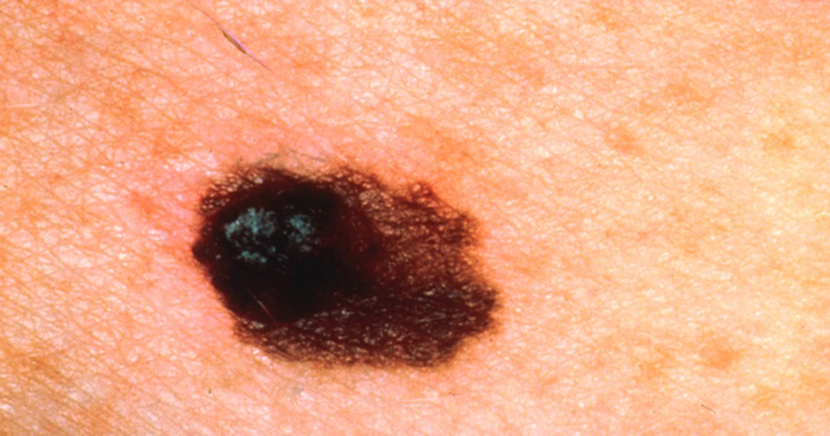
Table of contents
- Why melanoma requires a team
- Diagnosis: dermoscopy, excision and pathology report
- Staging: what the T category decides
- Sentinel lymph node and the end of routine dissection
- Decisions that must come before surgery
- Advanced disease: biomarker and treatment choice
- Immune-mediated toxicity: who does what
- Follow-up, older adults and the limits of the tools
- How NEXO helps to train this reasoning
- Frequently asked questions
- Final checklist
- References
A 58-year-old woman comes to dermatology because of a dark spot on her back that her husband noticed had changed color over the past few months. The lesion has irregular borders, two shades of brown and a bluish spot in the center. Dermoscopy reinforces the suspicion, and complete excision with a narrow margin confirms cutaneous melanoma with a Breslow thickness of 2.4 mm, without ulceration. Still in the office, she asks whether she will need to "remove all the lymph nodes", whether she will have chemotherapy and who will take care of her from now on. The scene is fictional and was written for teaching.
This text uses the case as a setting for clinical reasoning for students and health professionals. The topic is not an isolated study, but the organization of care: how dermatology, surgery, pathology, medical oncology, radiology and radiation oncology coordinate the diagnosis, staging and treatment of melanoma, which decisions need to be discussed before surgery and how to set up follow-up for immune-mediated toxicities. The text does not replace your service's protocol or your country's guideline. If you play the case of the day on NEXO, you will recognize the challenge: each clue changes the next step, and the order of the steps matters.
Key points
- The diagnosis starts in dermatology, with dermoscopy and complete excision with a narrow margin, before any wide surgery.
- Breslow thickness and ulceration define the T category, and the T category defines who discusses the sentinel lymph node biopsy.
- With a positive sentinel lymph node, immediate complete lymph node dissection did not increase melanoma-specific survival in MSLT-II.
- In stage III disease with a clinically detectable lymph node, the conversation about systemic treatment before surgery needs to happen before scheduling the operation.
- Immunotherapy requires a toxicity plan shared between oncology, nursing, the emergency department and the organ specialties.
- Follow-up is individual, with a complete skin examination preferably performed by a dermatologist.
Why melanoma requires a team
Cutaneous melanoma is a tumor in which the specialist who sees the lesion first is almost never the same one who decides on systemic treatment. Dermatology makes the suspicion, performs dermoscopy and often the diagnostic excision. Pathology measures the thickness, describes ulceration, mitoses and margins, and in metastatic disease tests for the BRAF mutation. Surgical oncology performs the margin widening and the sentinel lymph node biopsy, with support from nuclear medicine for lymphatic mapping. Radiology stages and follows up. Medical oncology leads adjuvant, neoadjuvant or advanced disease therapy. Radiation oncology comes in for specific scenarios, such as brain metastasis or palliation.
When each one decides alone, three typical failures arise. The first is margin widening performed before sentinel lymph node mapping, which can alter lymphatic drainage and harm the procedure. The second is operating on bulky lymph node disease without first discussing preoperative systemic treatment, missing a window that today has randomized evidence. The third is treating immunotherapy toxicity late, because the patient went to an emergency department that did not know what he was receiving.
The 2025 ESMO guideline for cutaneous melanoma recommends, at several points, that treatment be discussed at a multidisciplinary tumor meeting, especially in patients at high risk of distant metastasis. That is the thread running through this text: not a single algorithm, but a sequence of decisions with a clear owner.
What the multidisciplinary meeting is
The multidisciplinary tumor board is the periodic meeting in which specialists review imaging, pathology report and patient context together, and record a recommendation. In melanoma, it usually brings together dermatology, surgery, pathology, medical oncology, radiology, radiation oncology, nuclear medicine and nursing. The value lies less in the meeting itself and more in the record: who will do what, in what order and with which criterion to change the plan.
Who participates and in what role
| Specialty | Main role in melanoma | Moment when it is decisive |
|---|---|---|
| Dermatology | Suspicion, dermoscopy, diagnostic excision, skin examination at follow-up | Diagnosis and follow-up |
| Pathology | Breslow, ulceration, margins, BRAF V600 testing, pathological response after neoadjuvant therapy | Staging and after surgery |
| Surgical oncology | Wide local excision, sentinel lymph node, lymph node or metastasis resection | Localized and regional disease |
| Nuclear medicine | Lymphoscintigraphy and sentinel lymph node localization | Before surgery |
| Medical oncology | Adjuvant therapy, neoadjuvant therapy, advanced disease, toxicity management | Stage III and IV, some stage II |
| Radiology | Staging, lymph node ultrasound, response and follow-up | The entire pathway |
| Radiation oncology | Brain metastasis, palliation, selected adjuvant scenarios | Advanced disease |
| Nursing and organ specialties | Education, symptom triage, management of immune-mediated events | During and after immunotherapy |
Diagnosis: dermoscopy, excision and pathology report
In the scenario, the first correct decision was dermatology's: not to perform a wide excision straight away. The ESMO guideline recommends dermoscopy by an experienced physician, because it increases diagnostic accuracy, and recommends that the diagnosis be based on complete full-thickness excision with a minimal margin of clinically normal skin. In large lesions, where complete excision would require reconstruction, a biopsy may be performed. These recommendations are in the full text of the ESMO guideline.


Narrow excision has a logic of sequence. It gives the pathologist the entire lesion, which allows measuring the maximum thickness, and preserves lymphatic drainage for possible sentinel lymph node mapping. If the first surgery were already a 2 cm wide excision with flap rotation, subsequent mapping could be less reliable.
What the pathology report must include
The pathology report is the piece that feeds all subsequent decisions. The team should check whether it includes the items below before discussing the case.
- Breslow thickness, recorded to the nearest tenth of a millimeter, as required by the eighth edition of the AJCC.
- Presence or absence of ulceration.
- Status of the lateral and deep margins.
- Mitotic rate, which is no longer part of the T category but remains useful prognostic information.
- Histological subtype and presence of microsatellitosis, when present.
- In metastatic disease, result of BRAF V600 testing, preferably on the metastasis.
The eighth edition of the AJCC began requiring thickness rounded to the nearest tenth of a millimeter, rather than to the nearest hundredth, according to the review by Gershenwald and colleagues in CA Cancer J Clin. It seems like a detail of the report, but this decimal place decides, for example, whether a 0.8 mm tumor is T1a or T1b.
Staging: what the T category decides
The T category of melanoma depends on thickness and ulceration. In the eighth edition, T1a became a tumor less than 0.8 mm without ulceration, and T1b began to include tumors of 0.8 to 1.0 mm, with or without ulceration, and tumors less than 0.8 mm that are ulcerated. Mitotic rate is no longer a criterion for T. These changes are described in the publication by the AJCC committee.
In the scenario, 2.4 mm without ulceration corresponds to T3a. This has an immediate consequence: the patient is a candidate for sentinel lymph node biopsy. ESMO recommends sentinel lymph node biopsy for all patients with clinically negative lymph nodes and tumors T2a or higher, that is, above 1.0 mm, and recommends discussing it with T1b patients, according to the 2025 guideline.
Wide excision margins
Margin expansion also follows thickness. ESMO recommends margins of 0.5 cm for melanoma in situ, 1 cm for tumors up to 2 mm and 2 cm for tumors above 2 mm, in the guideline text. In the scenario, the planned margin is 2 cm, performed in the same surgical session as the sentinel lymph node.
Imaging tests in staging
Not every patient needs a whole-body CT scan at diagnosis. The intensity of imaging follows the stage, and the decision to order CT, PET or brain MRI should be made together with oncology and radiology, according to the local guideline. The reasoning point is to avoid two opposite errors: ordering too much imaging in thin disease, with incidental findings that generate anxiety and procedures, and ordering too little imaging in thick or lymph node disease, missing a metastasis that would change the whole plan.
Sentinel lymph node and the end of routine dissection
Sentinel lymph node biopsy is a staging procedure. The final MSLT-I report, with 2001 patients randomized between wide excision with lymph node observation and wide excision with sentinel lymph node biopsy, showed no difference in melanoma-specific survival at 10 years in the overall population. Among tumors of intermediate thickness, from 1.20 to 3.50 mm, 10-year disease-free survival was 71.3 in every 100 in the biopsy group versus 64.7 in every 100 in the observation group, in the same report.
The same study showed the prognostic weight of the result: among tumors of intermediate thickness, 10-year melanoma-specific survival was 62.1 in every 100 with lymph node metastasis and 85.1 in every 100 without metastasis, according to MSLT-I. That is why the sentinel lymph node informs the adjuvant treatment decision: it separates patients with very different risks.
What MSLT-II changed
For years, a positive sentinel lymph node led to complete lymph node dissection. MSLT-II randomized patients with a positive sentinel node between immediate complete dissection and observation with ultrasound. Immediate dissection did not increase melanoma-specific survival: at 3 years, it was 86 in every 100 in both groups in the per-protocol analysis, in the NEJM article. Disease-free survival was slightly higher with dissection, at the cost of more regional control and more morbidity, such as lymphedema.
The practical consequence is in the guideline: ESMO does not recommend complete lymph node dissection for patients with a positive sentinel lymph node, but standard follow-up, with or without systemic therapy depending on the stage, according to the 2025 guideline. This shifts the center of the decision from surgery to the multidisciplinary meeting, because the next step is no longer another operation but the conversation about adjuvant therapy and imaging surveillance.


Observation is not abandonment
Choosing observation instead of dissection does not mean leaving the patient without follow-up. In MSLT-II, the observed group had periodic lymph node ultrasound. In practice, this requires a radiology service that knows how to examine the lymph node basin and a workflow that brings the patient back quickly if a suspicious lymph node appears. This is a task for the team, not for a single specialist.
Decisions that must come before surgery
Dr. Alexei's central question for this tumor is which decisions should be discussed before surgery. The answer changed with the studies of perioperative systemic treatment in stage III disease with clinically detectable lymph node.
In the SWOG S1801 study, phase 2, patients with resectable stage IIIB to IVC melanoma and measurable disease were randomized between three doses of pembrolizumab before surgery, followed by the operation and 15 adjuvant doses, or surgery followed by pembrolizumab 200 mg intravenously every 3 weeks, for a total of 18 doses. In the data cutoff analysis, 2-year event-free survival was 72 in 100 in the neoadjuvant plus adjuvant group and 49 in 100 in the adjuvant-only group, according to the NEJM article. The dose cited is the one in the study protocol; the dose and duration in each country follow the label and the local guideline.
ESMO also describes the NADINA study, phase 3, which compared two doses of nivolumab plus ipilimumab before surgery against adjuvant nivolumab in 423 patients with resectable stage III melanoma with biopsy-confirmed lymph node involvement. In that design, treatment after surgery depended on the pathological response. NADINA has its own post in the series and is not detailed here.
Why this is a team decision
Preoperative treatment changes everyone's schedule. Surgery has to reserve the date after the planned doses. Pathology has to know how to measure pathological response in the index lymph node and in the specimen. Radiology needs baseline imaging and a reassessment. Oncology needs a plan for the patient who has toxicity and cannot undergo surgery on time. A Delphi consensus from an Irish cancer center on the implementation of neoadjuvant immunotherapy in stage III found consensus on most statements, but not on three points: the use of immunotherapy to make an unresectable disease resectable, adjuvant therapy adapted to response, and the ideal extent of lymph node resection.
What to discuss before scheduling the operation
- Is the lymph node disease clinically detectable and confirmed by biopsy, or only microscopic in the sentinel node?
- Is there distant disease or brain metastasis that changes the goal of treatment?
- Does the patient have a contraindication to immunotherapy, such as active severe autoimmune disease or a solid organ transplant?
- What is the surgery date if neoadjuvant treatment is chosen, and who reassesses before it?
- How will pathology describe the response, and what does that response change afterward?
- Who is the patient's contact if a symptom appears between doses?
Advanced disease: biomarker and treatment choice
In metastatic disease, the first laboratory question is the BRAF V600 mutation. ESMO recommends testing for it preferably in the metastasis or, failing that, in the primary tumor, and accepts circulating tumor DNA as an alternative when there is no tissue, according to the 2025 guideline. The same guideline prefers enrollment in a clinical trial whenever possible.
The BRAF result opens the possibility of targeted therapy with a BRAF inhibitor plus a MEK inhibitor, but does not require using it first. Immunotherapy with PD-1 blockade, alone or combined with CTLA-4 blockade, has long-term data. In the final 10-year analysis of CheckMate 067, with 945 previously untreated patients, median overall survival was 71.9 months with nivolumab plus ipilimumab, 36.9 months with nivolumab and 19.9 months with ipilimumab. These numbers show that some patients live many years, but they do not justify calling the treatment a cure: each patient's curve depends on response and toxicity.
Brain metastasis and radiotherapy
Brain metastasis changes the makeup of the team. Neurosurgery and radiotherapy join in, and the decision on radiosurgery, surgery or systemic treatment first depends on the number of lesions, the symptoms and the use of corticosteroids. This is one of the moments when the multidisciplinary meeting prevents delay: the patient does not need to go through three successive appointments to hear three separate opinions.
What the team records before the first cycle
Before the first cycle, the team records the BRAF result and where the sample came from, performance status, comorbidities and prior autoimmune diseases. It also records the baseline thyroid, glucose, liver and kidney tests requested by the service protocol, the goal of treatment explained to the patient without a promise of cure, and the contact channel for symptoms, with written guidance on when to seek care.
Immune-mediated toxicity: who does what
Immune checkpoint inhibitors can inflame almost any organ: skin, intestine, liver, lung, endocrine glands, joints, nerves and heart. That is why the ASCO guideline on immune-mediated adverse events was written by a panel that brought together oncology, dermatology, gastroenterology, rheumatology, pulmonology, endocrinology, urology, neurology, hematology, emergency medicine, nursing and patient representatives. The makeup of the panel already describes the team the patient needs.
The guideline sets out a general logic by grade. In grade 1 toxicity, treatment generally continues with monitoring, with neurological, hematological and cardiac exceptions. In most grade 2 toxicities, the inhibitor is held and can be resumed when symptoms return to grade 1 or less, and a corticosteroid can be given at a starting dose of 0.5 to 1 mg/kg/day of prednisone or equivalent. In grade 3, the inhibitor is held and a high-dose corticosteroid is started, prednisone or methylprednisolone at 1 to 2 mg/kg/day, with tapering over at least 4 to 6 weeks, according to the full ASCO text. Refractory cases may require infliximab or another immunosuppressant, per the same guideline.
Where care usually fails
The most common mistake is not one of pharmacological knowledge but of communication. The patient with diarrhea on a Sunday goes to the nearest emergency department, which treats it as viral gastroenteritis and sends them home. The immune-mediated colitis progresses. To avoid this, the team needs four things: a card or record stating what the patient is receiving, a contact line with oncology, a protocol agreed with the emergency department and a fast path to the organ specialty.
Roles in managing toxicity
- Oncology nursing: education before the first cycle and telephone symptom screening.
- Clinical oncology: grading the event, holding or resuming treatment and starting the corticosteroid.
- Emergency department: recognizing the patient on immunotherapy and contacting oncology before discharge.
- Organ specialties: endoscopy in colitis, bronchoscopy in pneumonitis, endocrinology in hypophysitis and thyroiditis, cardiology in myocarditis.
- Pharmacy: interaction of prolonged corticosteroid use with other drugs and prophylaxis when indicated.
Follow-up, older adults and the limits of the tools
Follow-up after treatment has no single schedule. ESMO states that there is no consensus on the ideal regimen or on the usefulness of imaging and blood tests in resected melanoma, that national guidelines should be consulted and that the schedule should be adjusted to each patient, considering stage, risk and personal needs. The same guideline recommends a skin examination, preferably by a dermatologist, to detect recurrence and new skin tumors, especially second melanomas, in the 2025 text.


The older patient
Melanoma weighs more heavily on the older population, and age changes the conversation. In a single-institution study of 240 patients with stage III melanoma, those aged 75 or older received less adjuvant immunotherapy than younger patients, and among those who did not receive it, 58 in 100 declined treatment. In that same study, advanced age was not associated with more toxicity, interruption or discontinuation of treatment, while adjuvant immunotherapy was associated with lower recurrence. The authors call for a better understanding of this difference in use and for listening to the patients' perspective. In practice, the decision should be guided by the tumor, physiological reserve, function, comorbidities and preferences, and not by chronological age alone.
Digital tools in the meeting
Language models are starting to be tested as decision support. A retrospective single-center study from Bosnia and Herzegovina compared recommendations from four models with the actual decisions of the melanoma multidisciplinary meeting in 151 patients, assessed by oncologists. Performance varied between the models, with statistically significant differences. For the student, the lesson is about method: the tool was measured against the team's decision, not the other way around. Responsibility for the recommendation remains with the specialists who know the patient.
Reasoning errors the team helps to avoid
- Premature closure: treating the lesion as an atypical nevus and postponing excision.
- Anchoring on the first report: not reviewing the slide when the thickness does not seem to match the clinical picture.
- Action bias: ordering lymph node dissection out of habit, after MSLT-II.
- Omission bias: not discussing neoadjuvant therapy because surgery was already scheduled.
- Wrong attribution of a symptom: treating diarrhea, cough or fatigue as a trivial cause in a patient on immunotherapy.
These biases appear in any field. The post on implicit bias linked to gender, race and age helps to understand why the older patient receives less treatment than they could, and the text on overdiagnosis helps to think about excess imaging in follow-up. Another example of immunotherapy with a toxicity plan appears in the post on enfortumab vedotin and pembrolizumab in urothelial cancer.
How NEXO helps to train this reasoning
NEXO works with fictional cases with a real clinical presentation, in which the clues arrive one at a time, up to six clues. This format trains the same skill that the multidisciplinary meeting requires: deciding what can already be concluded with what you have and what is still missing before the next step. In melanoma, this means not jumping from suspicion to wide surgery, not jumping from a positive sentinel node to dissection and not jumping from a nonspecific symptom to the trivial explanation.
If you like to study the mechanisms behind the clues, the Researcher area organizes deeper study. To close diagnoses with fewer clues, the Closer profile shows how to measure your own accuracy. Case curation is in Curator, and those who produce medical content find the program in Creators. The limits of use are in /economia. Other blog texts help to train the sequence of decisions, such as differential diagnosis with clues and interprofessional clinical reasoning.
Frequently asked questions
Does every melanoma need sentinel lymph node biopsy?
No. ESMO recommends biopsy for tumors above 1.0 mm with clinically negative lymph nodes and recommends discussing it in T1b tumors. In melanoma in situ and T1a, it is generally not indicated.
If the sentinel node is positive, will I have to remove all the nodes?
In most patients, no. MSLT-II showed no melanoma-specific survival gain with immediate complete dissection, and ESMO does not recommend it routinely. Follow-up then includes lymph node ultrasound, and the team discusses systemic therapy according to stage.
Why discuss treatment before surgery?
Because, in stage III disease with a clinically detectable lymph node, there is randomized evidence that giving immunotherapy before the operation improves event-free survival compared with giving it only afterward. That option only exists if it is discussed before surgery is performed.
Who treats immunotherapy toxicity?
Oncology coordinates, but management involves nursing, the emergency department and the specialties of the affected organ. The ASCO guideline advises suspending treatment and using corticosteroids according to grade.
Does immunotherapy cure metastatic melanoma?
It prolongs the lives of many patients, and some of them live more than 10 years, as CheckMate 067 shows. Even so, the text avoids calling this a cure, because there is no way to guarantee that result for an individual patient.
Does advanced age prevent treatment?
Not by itself. The decision should consider physiological reserve, function, cognition, comorbidities, nutrition and preferences, preferably with a geriatric assessment.
Final checklist
- Suspicious lesion: dermoscopy and complete excision with a narrow margin before any wider excision.
- Report: Breslow thickness to the nearest tenth of a millimeter, ulceration, margins and, in metastatic disease, BRAF V600.
- T category defined and sentinel lymph node indication decided with the patient.
- Margin expansion according to thickness, at the same time as the sentinel node.
- Positive sentinel node: discussion at a multidisciplinary meeting, without routine complete dissection.
- Clinically detectable lymph node disease: neoadjuvant therapy discussed before scheduling surgery.
- Immunotherapy: patient education, a contact channel and a protocol shared with the emergency department.
Play the case of the day on NEXO and practice deciding with the clues in the right order.
References
- Amaral T, Ottaviano M, Arance A, et al. Cutaneous melanoma: ESMO Clinical Practice Guideline for diagnosis, treatment and follow-up. Ann Oncol. 2025. PMID 39550033.
- Babic B, Umihanic S, Osmanovic H, et al. Clinical evaluation of large language model recommendations in melanoma: comparison with multidisciplinary tumor board decisions in a real-world cohort. Front Oncol. 2026. PMID 42444798.
- Brahmer JR, Lacchetti C, Schneider BJ, et al. Management of Immune-Related Adverse Events in Patients Treated With Immune Checkpoint Inhibitor Therapy: American Society of Clinical Oncology Clinical Practice Guideline. J Clin Oncol. 2018. PMID 29442540.
- Cronin C, Martin F, Martin-Smith JD, et al. Implementation of neoadjuvant immunotherapy in stage III melanoma: a modified Delphi consensus study in a European-accredited cancer center in Ireland. Oncologist. 2025. PMID 40857557.
- Bhatty MA, Chakraborty NN, Zablonski KG, et al. The Use and Utility of Adjuvant Checkpoint Inhibitors in Elderly Patients with Melanoma: A Single Institution Experience. Cancers (Basel). 2026. PMID 42352428.
- Faries MB, Thompson JF, Cochran AJ, et al. Completion Dissection or Observation for Sentinel-Node Metastasis in Melanoma. N Engl J Med. 2017. PMID 28591523.
- Gershenwald JE, Scolyer RA, Hess KR, et al. Melanoma staging: Evidence-based changes in the American Joint Committee on Cancer eighth edition cancer staging manual. CA Cancer J Clin. 2017. PMID 29028110.
- Morton DL, Thompson JF, Cochran AJ, et al. Final trial report of sentinel-node biopsy versus nodal observation in melanoma. N Engl J Med. 2014. PMID 24521106.
- Patel SP, Othus M, Chen Y, et al. Neoadjuvant-Adjuvant or Adjuvant-Only Pembrolizumab in Advanced Melanoma. N Engl J Med. 2023. PMID 36856617.
- Wolchok JD, Chiarion-Sileni V, Rutkowski P, et al. Final, 10-Year Outcomes with Nivolumab plus Ipilimumab in Advanced Melanoma. N Engl J Med. 2025. PMID 39282897.