Phlebitis and catheter infection: when access is the focus
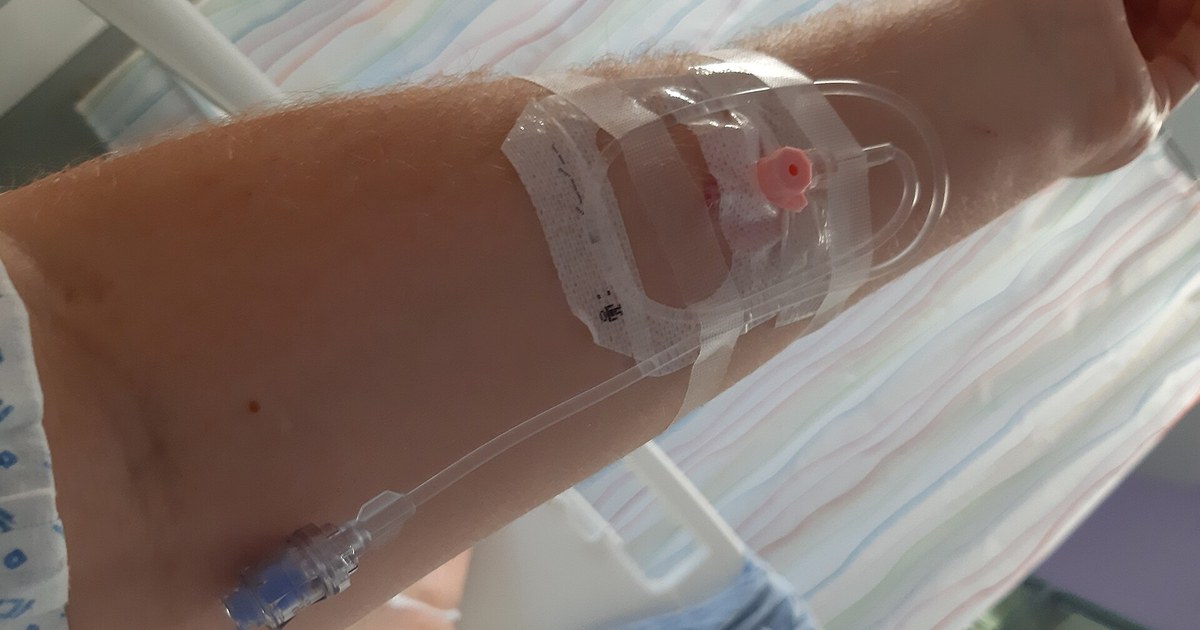
Table of contents
A 67-year-old woman has been in hospital for five days with pneumonia. She has been taking the antibiotic by mouth since yesterday, but the peripheral venous catheter in her forearm is still there, closed, unused for 48 hours. Nobody remembers who decided to keep it "just in case". At the morning handover, the patient has a fever of 38.6 degrees and chills. The nurse uncovers the dressing: the skin around the insertion point is red and warm, there is a hardened cord running up the vein for a few centimeters and a drop of yellowish discharge at the opening. The scene is fictional and was written for teaching.
This text uses the case as a setting for clinical reasoning in nursing. The fever looks like a relapse of the pneumonia, and the catheter looks like a detail. The correct reading reverses the order: the forgotten line has become the most likely focus, and the germ that matters most in this situation is Staphylococcus aureus. The text does not replace your institution's protocol or the medical prescription. If you play the case of the day on NEXO, you will recognize the challenge: look at the device before blaming the underlying disease.
Key points
- Phlebitis is common: in intensive care, it appeared in about 1 in every 13 peripheral catheters in a multicenter study.
- Pain, hardening, redness or discharge at the insertion point mean the peripheral catheter should be removed.
- Discharge at the insertion point should go for Gram stain and culture.
- Bloodstream infection from a peripheral catheter is rare, but it can be serious, especially when the germ is Staphylococcus aureus.
- An unused catheter is risk without benefit: the daily question is whether it is still needed.
- The site of a line that has already been removed also needs to be looked at in the following days.
Why this is a reasoning setting
The peripheral venous catheter is the most used invasive device in hospital, and that is exactly why it is easy to forget. In a prospective study of 842 catheters in adults, 141 of 359 patients had at least one complication, and phlebitis came first on the list, present in 17.6 percent of catheters, followed by pain, leakage, dislodgement, extravasation and obstruction (Abolfotouh and colleagues, 2014). In a Portuguese cohort, phlebitis and infiltration rates were 43.2 and 59.7 per 1,000 catheter-days, and most catheters with these vascular injuries were removed within the first 24 hours (Braga and colleagues, 2018).
The setting is good for training because the answer depends on putting together clues that sit in different places: the fever is on the vital signs chart, the catheter is under the dressing, and the information that it has been idle for two days is in the prescription. The post on signs, symptoms and history shows how to read what the patient brings, and the post on clinical deterioration on the ward shows how fever and the other signs fit into an alert score.
The cost of delay
In a Japanese series of 62 patients with peripheral catheter-associated bloodstream infection, the mean time between catheter insertion and diagnosis of bacteremia was 6 days, and 8 patients (12.9 percent) died within 30 days of the positive blood culture (Sato and colleagues, 2017). Those who died had a higher proportion of Staphylococcus aureus infection, with an odds ratio of 8.33 (Sato and colleagues, 2017). The authors report cases that required intensive care and prolonged antibiotics for distant complications (Sato and colleagues, 2017).
What the scene teaches
The scene trains four habits. The first is to examine the insertion point in every patient with a new fever. The second is to ask every day whether the catheter is still needed. The third is to know what to do with the discharge and with the catheter before throwing them in the bin. The fourth is to recognize when the local infection may have reached the blood.
Phlebitis: chemical, mechanical or infectious
Phlebitis is inflammation of the vein wall. In peripheral access, it is usually described in three groups, and the distinction matters because it changes what to expect from the course. The scoping review by Guanche-Sicilia and colleagues, done from the nursing point of view, notes that phlebitis shows up as redness, swelling along the vein, pain, warmth and fever, and that assessment scales help standardize the record (Guanche-Sicilia and colleagues, 2021).
Chemical
Chemical phlebitis comes from what runs inside the vein. In 3,429 catheters from 23 Japanese intensive care units, the only factors that increased the risk of phlebitis in the multivariate analysis were intravenous drugs; among 26 assessed, four were associated with more phlebitis: nicardipine, noradrenaline, amiodarone and levetiracetam (Yasuda and colleagues, 2022). Standardized drug administration measures in the unit were associated with lower risk (Yasuda and colleagues, 2022). In the Portuguese cohort, piperacillin with tazobactam was associated with infiltration (Braga and colleagues, 2018).
Mechanical
Mechanical phlebitis comes from the catheter rubbing against the vein wall: a catheter too thick for the vein, loose fixation, a flexion site. In the Japanese study, inserting in the arm instead of the forearm was associated with lower risk of phlebitis (Yasuda and colleagues, 2022). In the study by Abolfotouh and colleagues, female sex, insertion in the forearm or arm and drug infusion predicted phlebitis (Abolfotouh and colleagues, 2014).
Infectious
Infectious phlebitis is the one that worries most. It can stay limited to the site or reach the bloodstream. Signs such as purulent discharge, a very painful cord, fever and chills push the reasoning toward infection. In the case in the scene, the yellowish discharge at the puncture site and the fever with chills already take the picture out of the "simple chemical phlebitis" group.
| Type | Typical clue | What to think |
|---|---|---|
| Chemical | Appears during infusion of an irritating drug | Review the drug, the dilution and the route |
| Mechanical | Catheter at a flexion site, loose fixation | Review the site, the gauge and the fixation |
| Infectious | Discharge, fever, chills, worsening after removal | Remove it, culture the discharge, think of bacteremia |
Table sources: (Guanche-Sicilia and colleagues, 2021), (Yasuda and colleagues, 2022), (Braga and colleagues, 2018) and (Mermel and colleagues, IDSA, 2009). The division is didactic; in practice the mechanisms add up.
Who is at higher risk
The frequency of phlebitis varies widely between studies, because definitions and scales change. In the AMOR-VENUS multicenter study, with 2,741 patients and 7,118 peripheral catheters in intensive care, phlebitis occurred in 7.5 percent of catheters and in 12.9 percent of patients; most catheters were removed soon after diagnosis, grade 1 was the most common and grade 4 the rarest (Yasuda and colleagues, 2021). Catheter failure, adding up all causes, reached 21 percent (Yasuda and colleagues, 2021).
Factors linked to the patient and to the hospital stay
In the Portuguese cohort, length of stay and number of catheters inserted were risk factors for phlebitis (Braga and colleagues, 2018). That makes sense: each new puncture is a new entry point and a new insult to the vein. Patients with difficult veins accumulate attempts and accesses in unfavorable sites.
Factors linked to care
In an analysis of catheter tips in hospitalized adults, 41.8 percent of the catheters studied ended in failure, and 5.8 percent of the cultured tips grew organisms, mostly Staphylococcus epidermidis; more than half of the positive cultures came from patients with local signs consistent with catheter-related infection (Blanco-Mavillard and colleagues, 2019). Most episodes responded well to catheter removal and management (Blanco-Mavillard and colleagues, 2019). The finding reminds us that a tip culture alone does not diagnose infection; it must be read together with the clinical picture.
The idle catheter
A catheter with no use does not get the same attention as a catheter with an infusion running. Nobody opens the dressing to check a line that is not being used. In the case in the scene, 48 hours without use mean 48 hours of risk with no benefit. The question that prevents the problem is simple and daily: is this catheter still needed?
How to assess the access at the bedside
Assessing the access site is not just checking whether it "is working". It means looking, touching and asking, every shift and whenever there is a new fever.
Scales
There are several scales for grading phlebitis, such as the Visual Infusion Phlebitis, the phlebitis scale and the Maddox scale (Guanche-Sicilia et al., 2021). Sato et al. recommend examining the insertion site every day and using tools such as the Visual Infusion Phlebitis to keep the local infection from progressing to bacteremia (Sato et al., 2017). In a Delphi study with expert nurses in Spain, the items considered most important for assessing phlebitis were symptoms and observation, redness, the Maddox scale, induration, temperature and pain; in treatment, catheter removal was among the priority actions (Torné-Ruiz et al., 2024). The same authors found wide variation between services in assessment, treatment and follow-up (Torné-Ruiz et al., 2024). Use the scale adopted by your service, always the same one, so that progression is comparable between shifts.
What to observe and record
- Date and site of insertion, catheter gauge and who inserted it.
- Pain along the vein, even without visible redness.
- Redness, warmth, edema and a palpable cord, with its length in centimeters.
- Discharge at the puncture site: color, amount and whether it comes out spontaneously or on pressure.
- Fever, chills, tachycardia and a drop in blood pressure, which point to systemic involvement.
- Whether the catheter is in use, what is running through it and how long it has been unused.
Recording the measurement in centimeters shows whether the cord is growing. A photo in the medical record, when the service allows it, helps compare between shifts.


When phlebitis becomes bacteremia
Bloodstream infection from a peripheral catheter is far less frequent than phlebitis, but it is not negligible. In AMOR-VENUS, the rate was 0.8 percent (Yasuda et al., 2021). In the study by Blanco-Mavillard et al., the density was 0.76 per 1,000 admissions (Blanco-Mavillard et al., 2019).
Staphylococcus aureus and peripheral access
At an American academic center, among 445 episodes of Staphylococcus aureus bacteremia over two years, 34 (7.6 percent) were attributed to thrombophlebitis at the site of a peripheral catheter (Austin et al., 2016). Mortality at 90 days was comparable to that of other sources, and MRSA bacteremia linked to the peripheral catheter lasted longer (Austin et al., 2016). The most useful finding for nursing is another one: most of these infections were linked to old access sites, already removed, and not to the catheter in use and under surveillance (Austin et al., 2016). The authors argue that monitoring the site after removal should be a prevention priority (Austin et al., 2016).
How the bacteria reach the blood
There are two main routes. In the first, skin germs migrate along the catheter tract, from the puncture site inward, and form a biofilm on the device surface. In the second, contamination enters through the connector and the catheter lumen, with every manipulation. The biofilm protects the bacteria from the antibiotic and from the immune system, which explains why removing the device is a central part of treatment. An infected clot in the vein, suppurative thrombophlebitis, keeps the bacteremia going even after removal.
Signs of complication
The IDSA guideline lists situations that call for investigation of complications: bacteremia or fever that persist more than 3 days after adequate antibiotic therapy and catheter removal, especially when the organism is Staphylococcus aureus, and suspected suppurative thrombophlebitis (Mermel et al., IDSA, 2009). Transesophageal echocardiography is recommended when a shorter course than 4 to 6 weeks is considered in catheter-related Staphylococcus aureus bacteremia (Mermel et al., IDSA, 2009). Kouijzer et al. propose stratifying the risk of distant infection using patient factors, bacteremia factors (duration, short time to positivity, community acquisition, delay in treatment) and clinical course factors (persistent fever, unknown focus, signs of metastatic infection) (Kouijzer et al., 2023).
For nursing, this translates into concrete signs to report: fever that does not subside, new back or joint pain, a new murmur described by the physician, skin lesions, worsening of the cord after removal.
What to do at the bedside
The exact sequence depends on the local protocol. The actions below follow the IDSA guideline for peripheral catheters and the logic of not losing the focus.
The catheter
The guideline is direct: peripheral catheters with associated pain, induration, redness or drainage should be removed (Mermel et al., IDSA, 2009). In the patient in the scene, all these signs are present. Removal does not wait for the next scheduled change or the medical visit, and a new access, if still needed, goes to another site, preferably the other limb.
Drainage and cultures
Any drainage at the insertion site should be sent for Gram stain and routine culture, with fungal and mycobacterial cultures when the patient is immunocompromised (Mermel et al., IDSA, 2009). IDSA recommends collecting blood cultures before starting the antibiotic (Mermel et al., IDSA, 2009). For a central catheter, the recommendation is to collect paired samples, one through the catheter and one from a peripheral vein, labeling each bottle (Mermel et al., IDSA, 2009); for a peripheral catheter, blood cultures come from a puncture at another site. The catheter tip is cultured only when catheter-related infection is suspected, not routinely (Mermel et al., IDSA, 2009). Follow your service protocol on when and how to send the tip.
Communicating clearly
A structured message helps: "67-year-old patient, fifth day of hospitalization for pneumonia, already on oral antibiotics. Fever of 38.6 degrees with chills. Peripheral catheter in the left forearm unused for 48 hours, with redness, a 4-centimeter cord and purulent drainage. I removed the catheter and saved the drainage for culture. I request evaluation now and guidance on blood cultures and antibiotics." The sentence combines the likely focus, what has already been done and what is being requested.
The antibiotic
The choice and duration of the antibiotic are a medical decision, guided by the organism and the severity. When the organism is Staphylococcus aureus, IDSA recommends removing the catheter and treating for 4 to 6 weeks, with exceptions; a course of at least 14 days may be considered when the patient is not diabetic or immunosuppressed, the catheter has been removed, there is no intravascular prosthesis, there is no endocarditis or suppurative thrombophlebitis, and fever and bacteremia resolve within 72 hours of adequate antibiotic therapy (Mermel et al., IDSA, 2009). Practice varies widely around the world: in a survey of 2,031 physicians from 71 countries, treatment choices, the use of oral antibiotics and even the definition of persistent bacteremia changed by continent (Westgeest et al., 2023). The authors conclude that there is no international standard of care for this bacteremia (Westgeest et al., 2023).
Local measures
Compresses, ointments and limb elevation appear in reviews as measures for phlebitis, with heterogeneous evidence (Guanche-Sicilia et al., 2021), (Torné-Ruiz et al., 2024). They relieve discomfort, but they do not replace catheter removal or the investigation of infection.
Changing by routine or by clinical indication
For a long time, the rule was to change the peripheral catheter at a fixed interval, even without a problem. This rule was tested, and the result is not as simple as it seems.
What the studies showed
In an Australian randomized clinical trial with 755 patients, replacement based on clinical indication did not increase catheter failure due to phlebitis or infiltration compared with routine replacement every 3 days, and costs were lower (Webster et al., 2008). The phlebitis rate was low in both groups (4 and 3 percent) (Webster et al., 2008). A later meta-analysis, with 9 trials and 10,973 patients, found more phlebitis, occlusion and infiltration with replacement based on clinical indication, with no difference in local infection or in bloodstream infection (Chen et al., 2022). The authors suggest caution before abandoning routine replacement (Chen et al., 2022).
What this means in practice
Both strategies only work with surveillance. Replacement based on clinical indication assumes looking at the access every shift and removing it at the first sign. Routine replacement does not remove the need for that look, because infection can appear before the deadline. Neither one justifies keeping a catheter that is not being used. The replacement policy is a decision for the service; the role of those at the bedside is to examine, record and remove when there is a sign or when the catheter is no longer needed.
Prevention that depends on nursing
- Insert only when there is an indication and remove as soon as there is no longer one.
- Clean your hands before touching the catheter, the dressing or the connector.
- Disinfect the connector before each access, according to the protocol.
- Keep the dressing clean, dry and intact, and change it when it is not.
- Choose the gauge and site suited to the vein and to the infusion.
- Examine the site of removed accesses in the following days.
Sources for the list: (Mermel et al., IDSA, 2009), (Guanche-Sicilia et al., 2021) and (Austin et al., 2016). The details of antiseptics, dressings and intervals follow the service protocol.


Step-by-step reasoning in the case
It is worth retracing the case in order, as if the clues arrived one at a time.
First clue: fever on the fifth day
The first hypothesis is usually the disease that brought the patient in: the pneumonia did not respond. It is a reasonable hypothesis, but she is already on antibiotics and had been improving. New fever in the middle of a hospital stay calls for the list of hospital foci: catheters, urinary catheter, wound, the lung again, antibiotic-associated diarrhea, plus non-infectious causes such as thrombosis and drug reaction.
Second clue: the idle catheter
A device unused for 48 hours moves up the list of suspects. It should not have been there, and what should not be there tends not to be watched. The next step is to find out about the dressing.
Third clue: the insertion site
Redness, warmth, a hardened cord and purulent secretion confirm infectious phlebitis. The differential is now narrower: cellulitis around the access, infected superficial venous thrombosis, chemical phlebitis that coincided with another cause of fever. The secretion and the chills point to infection.
Fourth clue: what the chills say
Chills suggest bacteria passing into the blood. The question is no longer only "phlebitis?" and becomes "catheter-associated bacteremia, possibly due to Staphylococcus aureus?". This changes the urgency: remove, culture, communicate, prepare blood cultures before the antibiotic and watch for signs of complication in the following days.
Fifth clue: the course
If fever and positive blood cultures persist for more than 3 days after removal and the antibiotic, the team looks for complications such as suppurative thrombophlebitis and endocarditis (Mermel et al., IDSA, 2009). Nursing is who first sees the cord that grows, the new pain and the fever curve that does not fall.
| Hypothesis | For | Against | What decides |
|---|---|---|---|
| Pneumonia without response | Underlying disease, fever | Had been improving, already on oral antibiotic | Respiratory exam, imaging, course |
| Chemical phlebitis | Cord and pain along the tract | Catheter unused for 48 hours, purulent secretion | What was running through the catheter and when |
| Local infectious phlebitis | Secretion, redness, warmth | No systemic signs (present here) | Culture of the secretion |
| Catheter-associated bacteremia | Fever, chills, evident focus | Negative blood cultures | Blood cultures before the antibiotic |
| Superficial venous thrombosis | Hardened cord | Purulent secretion | Exam and, if indicated, ultrasound |
Table sources: (Mermel et al., IDSA, 2009), (Sato et al., 2017) and (Austin et al., 2016). The table organizes the reasoning; the diagnosis belongs to the team.
How NEXO helps train this reasoning
In NEXO, you try the diagnosis by searching for the ICD-10 code and receive proximity feedback at five levels, which shows whether the guess landed on the exact code, the same category, the same group, the same chapter or a different chapter. This format fits the theme of this text: pneumonia, phlebitis, vascular device-related infection, staphylococcal septicemia and endocarditis sit in different places in the classification, but they can be in the same patient on the same morning. The post about ICD-10 shows how to find the right code.
To use the case of the day as training, try three habits. Before the first guess, ask what devices the patient has. With each clue, ask whether it speaks of the underlying disease or of something the hospital added. After the result, write down the clue you left for later because it seemed small.
Three exercises with cases
List the devices. In each inpatient case, say which catheters, tubes and drains the patient has and for how long.
Separate local from systemic. Say which signs speak of infection at the insertion site and which speak of the blood.
Plan the surveillance. Say what you will watch in the days after catheter removal.
The post about differential diagnosis with clues shows how to organize hypotheses, the post about sepsis in nursing triage shows how to act when the infection already affects the whole body, the post about case-based learning shows how cases help fix these habits, and the post about diagnostic uncertainty discusses how to decide when the data do not yet add up.
The limits on using the game are described on the NEXO Economy page. If you research clinical reasoning teaching, the page for Researcher explains how to get in touch. Professionals who write or review cases find information on Curator and on Creators, and institutions can learn about the proposal on the partnerships page.
Frequently asked questions
Is every phlebitis an infection?
No. Phlebitis can be chemical or mechanical. Purulent discharge, fever and chills point to infection (Guanche-Sicilia et al., 2021), (Mermel et al., IDSA, 2009).
When should the peripheral catheter be removed?
When there is pain, hardening, redness or discharge at the site (Mermel et al., IDSA, 2009), and whenever it is no longer needed.
Should I send the discharge for culture?
Yes. IDSA recommends Gram stain and culture of any discharge at the insertion site (Mermel et al., IDSA, 2009).
Does changing every 3 days prevent infection?
Changing by clinical indication did not increase infection in the studies, but a meta-analysis found more phlebitis and infiltration without routine replacement (Webster et al., 2008), (Chen et al., 2022). Follow your service's policy and monitor the access.
Why look at an access that has already been removed?
Because many Staphylococcus aureus bacteremias linked to peripheral catheters came from old sites (Austin et al., 2016).
Final checklist
- I examined the insertion site in every patient with new fever.
- I asked whether the catheter was still needed and removed what was not.
- I graded the phlebitis with the service scale and measured the cord in centimeters.
- I removed the catheter with pain, hardening, redness or discharge.
- I sent the discharge for Gram stain and culture and remembered the blood cultures before the antibiotic.
- I communicated the probable focus, systemic signs and what had already been done.
- I watched fever, cord and signs of complication in the following days.
Want to practice reasoning with progressive clues? Play the case of the day.
References
- Abolfotouh MA, Salam M, Bani-Mustafa A, et al. Prospective study of incidence and predictors of peripheral intravenous catheter-induced complications. Ther Clin Risk Manag. 2014. PMID 25525365.
- Austin ED, Sullivan SB, Whittier S, et al. Peripheral Intravenous Catheter Placement Is an Underrecognized Source of Staphylococcus aureus Bloodstream Infection. Open Forum Infect Dis. 2016. PMID 27191005.
- Blanco-Mavillard I, Rodríguez-Calero MÁ, de Pedro-Gómez J, et al. Incidence of peripheral intravenous catheter failure among inpatients: variability between microbiological data and clinical signs and symptoms. Antimicrob Resist Infect Control. 2019. PMID 31367345.
- Braga LM, Parreira PM, Oliveira ASS, et al. Phlebitis and infiltration: vascular trauma associated with the peripheral venous catheter. Rev Lat Am Enfermagem. 2018. PMID 29791668.
- Chen CY, Chen WC, Chen JY, et al. Comparison of clinically indicated replacement and routine replacement of peripheral intravenous catheters: A systematic review and meta-analysis of randomized controlled trials. Front Med (Lausanne). 2022. PMID 36035414.
- Guanche-Sicilia A, Sánchez-Gómez MB, Castro-Peraza ME, et al. Prevention and Treatment of Phlebitis Secondary to the Insertion of a Peripheral Venous Catheter: A Scoping Review from a Nursing Perspective. Healthcare (Basel). 2021. PMID 34069674.
- Kouijzer IJE, Fowler VG, Ten Oever J. Redefining Staphylococcus aureus bacteremia: A structured approach guiding diagnostic and therapeutic management. J Infect. 2023. PMID 36370898.
- Mermel LA, Allon M, Bouza E, et al. Clinical practice guidelines for the diagnosis and management of intravascular catheter-related infection: 2009 Update by the Infectious Diseases Society of America. Clin Infect Dis. 2009. PMID 19489710.
- Sato A, Nakamura I, Fujita H, et al. Peripheral venous catheter-related bloodstream infection is associated with severe complications and potential death: a retrospective observational study. BMC Infect Dis. 2017. PMID 28623882.
- Torné-Ruiz A, Reguant M, Sanromà-Ortiz M, et al. Assessment, Treatment, and Follow-Up of Phlebitis Related to Peripheral Venous Catheterisation: A Delphi Study in Spain. Healthcare (Basel). 2024. PMID 38338263.
- Webster J, Clarke S, Paterson D, et al. Routine care of peripheral intravenous catheters versus clinically indicated replacement: randomised controlled trial. BMJ. 2008. PMID 18614482.
- Westgeest AC, Buis DTP, Sigaloff KCE, et al. Global Differences in the Management of Staphylococcus aureus Bacteremia: No International Standard of Care. Clin Infect Dis. 2023. PMID 37310693.
- Yasuda H, Yamamoto R, Hayashi Y, et al. Occurrence and incidence rate of peripheral intravascular catheter-related phlebitis and complications in critically ill patients: a prospective cohort study (AMOR-VENUS study). J Intensive Care. 2021. PMID 33407891.
- Yasuda H, Rickard CM, Marsh N, et al. Risk factors for peripheral intravascular catheter-related phlebitis in critically ill patients: analysis of 3429 catheters from 23 Japanese intensive care units. Ann Intensive Care. 2022. PMID 35394571.