Neck gunshot wound: hard signs and a zone-free approach
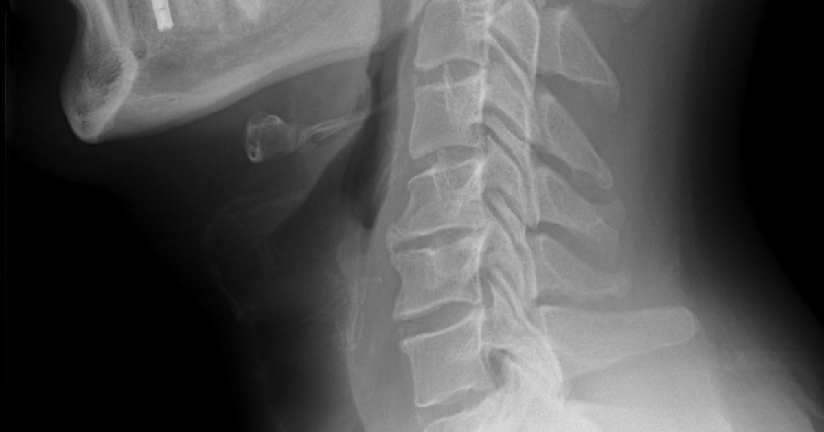
Table of contents
- Why the neck is such a dangerous region
- The neck zones and why they lost ground
- Airway first
- Bleeding control
- Hard signs and soft signs
- The role of CT angiography
- Esophagus and trachea, the injuries that slip through
- What the numbers show
- How NEXO helps train this reasoning
- Frequently asked questions
- Take-home messages
- References
The following scene is fictional and was written for teaching. A 34-year-old woman arrives at the emergency department with a gunshot wound to the left lateral side of the neck, just above the clavicle. She is conscious, speaks with a hoarse voice and occasionally spits blood. Her blood pressure is 112 over 70 mmHg and her saturation is 95 out of 100 on room air. There is a hematoma that does not appear to be growing and palpation reveals crepitus under the skin. There is no exit wound. The resident remembers the neck zones he studied and tries to classify the wound. The more experienced surgeon says the zone matters less than the signs and the imaging.
This text summarizes the current approach to gunshot wounds to the neck: the airway, hard and soft signs, the role of CT angiography and the shift from a zone-based approach to a sign-guided approach. The content is educational, aimed at students and health professionals, and does not replace local protocols or the surgical team's assessment.
Key points
- The neck packs the airway, vessels, esophagus, nerves and spine into a small space; the projectile does not follow anatomical planes and can injure structures far from its apparent path.
- Hard signs, such as expanding hematoma, severe active bleeding, refractory shock and airway obstruction, indicate immediate surgery.
- Soft signs, such as hemoptysis, dysphonia, dysphagia and subcutaneous emphysema, call for investigation, usually with CT angiography.
- CT angiography has become the initial test in the stable patient and helps define vascular injury and the projectile path.
- The internal injury lies in the same zone as the external wound in just over half of patients, which is why the zone-based approach has been falling out of use.
- The airway must be secured early when there is stridor, hoarseness, bubbling, tracheal deviation or extensive emphysema.
Why the neck is such a dangerous region
The neck brings together, within a few centimeters and with no bony protection in front, the larynx, trachea, pharynx, esophagus, carotid and vertebral arteries, jugular veins, cranial nerves, brachial plexus and cervical spinal cord. An Iranian review of 192 penetrating neck injuries starts precisely from this point: the region has a high density of vital structures in a small, unprotected area, which makes it one of the most vulnerable in the body (Mahmoodie and colleagues, 2012).
The mechanism also matters. In the Iranian series, most injuries were from sharp weapons, and the authors compare their findings with series in which the most common mechanism was gunshot (Mahmoodie and colleagues, 2012). Compared with a blade, the projectile does not follow anatomical planes, can deflect off bone and fragment, and the temporary cavity injures structures away from its path.
Gunshot wounds to the head and neck also have much higher in-hospital mortality than wounds in other regions. A national analysis of hospitalizations in the United States found mortality of 25.7 out of 100 in this group, against 2.7 out of 100 in other gunshot wounds (Menezes and colleagues, 2023). Part of this difference comes from the skull, but the neck contributes vascular and airway injuries that kill within minutes.
The neck zones and why they lost ground
For decades, the neck was divided into three zones. Zone I runs from the clavicles and the sternal notch to the cricoid cartilage. Zone II runs from the cricoid to the angle of the mandible. Zone III runs from the angle of the mandible to the base of the skull (Offiah and Hall, 2012). The same authors recall that, traditionally, zone I and III injuries were treated more conservatively because of the difficulty of surgical access. The division had surgical logic: zone II is easy to explore, while zones I and III require more complex approaches, such as sternotomy or a skull base approach.
The problem is that the external opening does not say where the internal injury is. In a South African study of 1,075 patients with penetrating neck injury, 298 had confirmed vascular or aerodigestive tract injury. In 176 of them, or 59.1 in every 100, the internal injury was in the same zone as the external wound. In 70 patients there was no correlation, and in 52 it could not be determined (Madsen and colleagues, 2020). The authors observed that all assessable patients with significant injury had suggestive physical signs or deep emphysema on imaging.
This finding supports the zone-free approach: instead of deciding by the location of the opening, the team decides by clinical signs and imaging. The same group proposes a zone-free flowchart, in which the unstable patient goes to the operating room and the stable patient is investigated according to signs and imaging (Madsen and colleagues, 2020). The zones remain useful for describing the wound and planning surgical access, but they no longer dictate management.
Airway first
The airway can be compromised by direct injury to the larynx or trachea, by compression from a hematoma, by blood in the oropharynx or by progressive edema. The review by Simpson and colleagues lists airway compromise or injury, apnea or respiratory failure, hypoxia and decreased level of consciousness as indications for intubation, and highlights that recognizing these indications early is crucial (Simpson and colleagues, 2021). Waiting for complete obstruction turns a difficult intubation into a surgical emergency.
A scoping review on the prehospital management of these injuries found that, in the studies analyzed, 67.2 in every 100 patients were intubated by rapid sequence with direct laryngoscopy, successfully in all of them (Simpson and colleagues, 2021). This does not mean intubation is simple, but rather that, in trained hands and with preparation, it is usually possible. The team must have an alternative plan ready, including a supraglottic device, a video laryngoscope and material for a surgical airway.
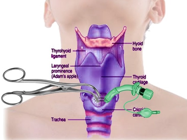
When oral intubation is not possible, cricothyroidotomy is the emergency surgical route. In extensive injuries of the cricoid or upper trachea, tracheostomy may be necessary. In open tracheal injuries, the tube is sometimes passed directly through the wound. A report of two Indian patients with open tracheal injury describes the airway difficulties caused by the combination of bleeding, blood aspiration, obstruction and intense sympathetic stimulation (Bhattacharya and colleagues, 2009).
The choice of location and timing also matters. Intubating in the emergency room, with the whole team and the material at hand, is usually safer than trying during transport or in the corridor. When the airway looks difficult, some services prefer to take the patient to the operating room and intubate there, with the surgeon ready for a surgical airway. Sedation must be done carefully, because muscle relaxation can make a hematoma collapse the airway the patient was maintaining with effort.
The signs that call for immediate attention to the airway are:
- Stridor or noisy breathing.
- Hoarseness or voice change.
- Air bubbling through the wound.
- Extensive or progressive subcutaneous emphysema.
- Tracheal deviation from a hematoma.
- Abundant blood in the oropharynx or hemoptysis.


Bleeding control
Neck bleeding can be external, visible in the wound, or internal, forming a hematoma that compresses the airway. Direct pressure with gauze is the classic initial measure, and the review by Simpson and colleagues recalls that simple dressings and direct compression were traditionally the basis of prehospital treatment (Simpson and colleagues, 2021). Hemostatic gauze has been studied in this context.
In the unstable patient with exsanguinating bleeding, one described option is inserting a Foley catheter into the wound and inflating the balloon to tamponade the bleeding while the patient is taken to the operating room (Madsen et al., 2020). The review by Simpson et al. also cites the Foley catheter among hemorrhage control measures (Simpson et al., 2021). It is a temporary measure that buys time until definitive control.
One precaution that is often forgotten is the cervical collar. The review by Simpson et al. cites an analysis of wounded soldiers that concluded the collar can hide signs of serious conditions, such as tracheal deviation, subcutaneous emphysema, expanding hematoma and diminished or absent carotid pulse (Simpson et al., 2021). In the review by Simpson et al., the odds ratio for death among patients who underwent spinal stabilization was 2.06 (Simpson et al., 2021). In penetrating wounds without neurologic deficit, unstable spinal injury is uncommon, and routine immobilization can hinder more than it helps.
Hemorrhage control is discussed in more detail in the text on tourniquet and the MARCH sequence. In the neck, the tourniquet does not apply, and direct compression, the balloon catheter and rapid transport take its place.
Hard signs and soft signs
The division into hard and soft signs is the basis of the modern approach. Hard signs indicate significant injury with high probability and generally take the patient straight to surgery. Soft signs raise suspicion and call for investigation. The review by Simpson et al. notes that hard signs occur in fewer than 10 out of every 100 patients (Simpson et al., 2021), which means most stable wounded patients go through investigation.
| Hard signs | Soft signs |
|---|---|
| Expanding or pulsatile hematoma | Hematoma that is not growing |
| Severe active bleeding | Hemoptysis, hematemesis or blood in the oropharynx |
| Shock that does not respond to volume | Dyspnea |
| Diminished or absent radial pulse | Dysphonia or dysphagia |
| Bruit or thrill | Subcutaneous or mediastinal air |
| Cerebral ischemia | Air leak from the chest drain |
| Airway obstruction | Focal neurologic deficit |
The table gathers the signs most cited in the reviews. The table adapted by Simpson et al. also includes air bubbling from the wound and massive emphysema among the hard signs, and proximity of the wound to the carotid or jugular, voice change and crepitus among the soft signs (Simpson et al., 2021). The lists vary a little between sources, but the logic is the same.
In the fictional opening scene, the patient has a hoarse voice, blood-streaked sputum and crepitus, three soft signs that suggest aerodigestive tract injury. She is stable, which allows investigation, but the hoarseness already calls for preparation to protect the airway. The text on diagnostic pause describes the habit of stopping and reviewing before closing the reasoning, useful precisely when the patient looks well.
The role of CT angiography
In the stable patient, cervical CT angiography has become the initial exam. An imaging review of penetrating trauma to the face and neck advocates CT angiography as the first-line exam (Offiah and Hall, 2012). A review of craniocervical vascular imaging describes CT angiography as a technique with superior sensitivity, specificity and accuracy for identifying vascular injury (Kee-Sampson et al., 2022).
The exam also shows the tract, which helps decide the next steps. When the tract passes far from critical structures, clinical observation is usually enough. When it passes near the pharynx, esophagus or trachea, investigation continues. The craniocervical vascular imaging review by Kee-Sampson et al. describes the findings of arterial injury on CT angiography, such as wall irregularity, occlusion, contrast extravasation, pseudoaneurysm and arteriovenous fistula (Kee-Sampson et al., 2022).
Doppler ultrasonography can help in some scenarios, but it depends heavily on the operator and assesses poorly the zones near the clavicle and the skull base. Plain radiography shows projectiles, fragments and air in the soft tissues, and can suggest airway or esophageal injury when there is deep emphysema, but it does not replace CT angiography.
Limits matter too. Projectile artifacts and metal fragments can impair the image near the metal. Small intimal injuries can go unnoticed. So doubtful findings may require conventional angiography, which also allows endovascular treatment in some cases.

Esophagus and trachea, the injuries that slip through
Pharyngeal and esophageal injuries are the ones that slip through most often. They usually show few signs in the first hours and, when untreated, progress to mediastinitis, with high mortality. The review by Offiah and Hall states that delayed diagnosis is the main factor for mortality associated with esophageal injury, estimated at about 20 in every 100, and that the main cause of death is mediastinitis (Offiah and Hall, 2012). The authors recommend contrast esophagography, endoscopy or both when clinical and radiological suspicion is high or when CT angiography already shows injury.
Laryngeal and tracheal injuries may present with hoarseness, emphysema, hemoptysis and air bubbling through the wound. Bronchoscopy or laryngoscopy helps confirm the injury and plan the repair. Small injuries can be treated conservatively, but extensive injuries require surgical repair and, sometimes, tracheostomy.
The order of investigation of the aerodigestive tract usually follows this reasoning:
- CT angiography to define the trajectory and vascular injury.
- If the trajectory passes near the esophagus or pharynx, contrast esophagography.
- If esophagography is not diagnostic, esophagoscopy.
- If there are signs of airway injury, laryngoscopy or bronchoscopy.
- Serial clinical observation, with reassessment of pain, fever, dysphagia and emphysema.
Sequelae can be permanent. Brachial plexus injury, dysphagia, vocal cord paralysis and airway stenosis requiring tracheostomy are among the consequences that can accompany the patient for life, and follow-up with speech therapy, otorhinolaryngology and rehabilitation is part of the treatment.


What the numbers show
Before the numbers, it is worth summarizing what the selective approach requires to work safely:
- A surgical team available to take the patient to the operating room at any time.
- CT angiography accessible without delay, with a report by someone who knows trauma.
- Access to esophagography, endoscopy and bronchoscopy when the trajectory indicates.
- Observation with repeated physical examination by professionals who know what to look for.
- A written protocol, so the decision does not depend on who is on duty.
Where these resources are lacking, surgical exploration may remain the safest option, and the decision should take the local context into account.
The shift from mandatory exploration to the selective approach shows in the numbers. In London, a trauma center compared its current experience with that of the 1980s: negative cervical exploration fell from 65 in every 100 in 1987 to 38 in every 100 in 2011. The authors concluded that selective management, based on the absence of instability and on imaging findings, reduces length of stay, surgical burden and costs, without increasing morbidity or mortality (Harris et al., 2012).
In São Paulo, a study of 161 patients, of whom 18.4 in every 100 had gunshot wounds, compared immediate exploration with selective exploration. Among the 130 in the selective group, 34 needed surgery after careful physical examination and tests guided by clinical indication. The selective group had a mean stay of 2 days and no deaths, and there was no statistical difference in mortality and complications between the groups (Teixeira et al., 2016).
In the Iranian series of 192 injuries, most from bladed weapons, zone II was the most affected, in 56.3 out of every 100 patients, and cervical exploration was positive in 84.4 out of every 100 explored (Mahmoodie et al., 2012). In a military registry of the Iraq and Afghanistan wars, of the 2,186 patients with penetrating cervical injury, 709 underwent cervical exploration, and 555 had a cervical vascular injury recorded (Breeze et al., 2020). Different settings produce different numbers, but the direction is the same: decide by the signs and by imaging, not by the map.
Damage control in the neck
A recent review on penetrating cervical trauma defines the injury as one that crosses the platysma muscle between the base of the skull and the sternum. It recalls that isolated cervical injuries account for up to 10 percent of trauma visits and that mortality can reach 10 percent (Loss et al., 2025). The same authors separate hard signs, such as expanding hematoma, severe active bleeding, shock unresponsive to volume, reduced or absent radial pulse, bruit or thrill, cerebral ischemia and airway obstruction, from soft signs, such as hemoptysis, hematemesis, dysphonia, dysphagia and subcutaneous air (Loss et al., 2025).
In the unstable patient, the authors describe a Foley catheter inserted into the wound and inflated to tamponade the bleeding until the operating room. The stated goal is to keep systolic pressure around 90 mmHg: lower pressures can worsen a cerebral infarct if there is cervical vessel injury, and high pressures can dislodge the clot (Loss et al., 2025). The review further stresses that blind intubation should not be attempted and that a surgical airway comes first when the anatomy is destroyed (Loss et al., 2025).
The World Society of Emergency Surgery review on face and neck trauma applies damage control principles to these patients. The immediate threat comes from the airway, compromised by bleeding, edema and loss of mandibular support. After securing the airway by orotracheal intubation, cricothyroidotomy or tracheostomy, direct compression and wound packing control almost all bleeding, except catastrophic cases (Krausz et al., 2015). The authors warn that attempting definitive repair in someone who already has major physiologic derangement tends to give poor results, and that damage control used without criteria exposes the patient to unnecessary procedures (Krausz et al., 2015).
The same review suggests dividing the neck structures into functional groups, such as airway, great vessels and digestive tract, so that assessment does not forget any of them (Krausz et al., 2015).
How NEXO helps train this reasoning
Training with fictional clinical cases helps recognize patterns before meeting them on shift, as long as each clue is read calmly and compared with the hypotheses that are still open. In cervical trauma cases, the clues may bring a hoarse voice, crepitus or an imaging finding that changes the reasoning. Reading each clue without clinging to the first hypothesis is the exercise described in the text on differential diagnosis with clues.
The texts on diagnostic uncertainty and clinical decision-making also help. To understand points and rewards, see the NEXO economy page. There are also tracks for those who research (Researcher), for those who review cases (Curator), for those who produce content (Creators) and for those who want to follow results (Closer).
Frequently asked questions
Do the neck zones still serve any purpose?
They serve to describe the wound and plan surgical access, since injuries at the base of the neck or near the base of the skull require different approaches. But the decision to operate or investigate depends on clinical signs and imaging, not on the external hole.
Does every gunshot wound to the neck need surgery?
No. Patients with hard signs go to surgery. Stable patients without hard signs are usually investigated with CT angiography and, depending on the trajectory, esophageal and airway studies. Many are treated without surgery.
When to intubate?
Early, when there is stridor, hoarseness, bubbling, tracheal deviation, extensive emphysema or abundant blood in the airway. Intubation requires preparation, with alternatives ready, including the surgical airway.
Why can the cervical collar get in the way?
Because it hides the neck. A growing hematoma, emphysema and tracheal deviation go unnoticed. In penetrating wounds without neurological deficit, unstable spine injury is uncommon, and immobilization should be used with judgment.
Why is esophageal injury such a concern?
Because it can show few signs at first and progress to mediastinitis, with high mortality. CT misses some of these injuries, so trajectories near the esophagus call for an esophagogram and, if needed, esophagoscopy.
What should be done with a wound that looks superficial?
Do not explore it with a finger, forceps or probe in the emergency room, because this can dislodge a clot and cause severe bleeding. Depth is assessed by clinical signs and imaging. If the platysma, the thin muscle just below the skin, was crossed, the workup follows the path described in this text.
How long should a stable patient with no findings be observed?
It depends on the local protocol, but observation usually lasts at least a few hours, with repeated examination of the airway, voice, swallowing, neck and pulses. Fever, pain on swallowing, new emphysema or worsening voice call for immediate reassessment.
Take-home messages
- Assess the airway first and intubate early when there are signs of threat.
- Compress external bleeding and consider a balloon catheter in the unstable patient.
- Use the cervical collar with judgment, without hiding the neck.
- Separate hard signs, which lead to surgery, from soft signs, which call for investigation.
- Perform CT angiography in the stable patient.
- Investigate the esophagus and airway when the trajectory passes near them.
- Decide by the signs and the imaging, not by the zone of the wound.
If you like reasoning from clues, the case of the day is waiting on the NEXO home page.
References
- Bhattacharya P, Mandal MC, Das S, et al. Airway management of two patients with penetrating neck trauma. Indian J Anaesth. 2009. PMID 20640146.
- Breeze J, Bowley DM, Combes JG, et al. Outcomes following penetrating neck injury during the Iraq and Afghanistan conflicts: A comparison of treatment at US and United Kingdom medical treatment facilities. J Trauma Acute Care Surg. 2020. PMID 32068717.
- Harris R, Olding C, Lacey C, et al. Changing incidence and management of penetrating neck injuries in the South East London trauma centre. Ann R Coll Surg Engl. 2012. PMID 22613301.
- Kee-Sampson JW, Gopireddy DR, Vulasala SSR, et al. Role of imaging in penetrating vascular injuries of the craniocervical region. J Clin Imaging Sci. 2022. PMID 36601604.
- Krausz AA, Krausz MM, Picetti E. Maxillofacial and neck trauma: a damage control approach. World J Emerg Surg. 2015. PMID 26157475.
- Loss L, Henry R, White A, et al. Penetrating neck trauma: a comprehensive review. Trauma Surg Acute Care Open. 2025. PMID 40166772.
- Madsen AS, Bruce JL, Oosthuizen GV, et al. Correlation between the level of the external wound and the internal injury in penetrating neck injury does not favour an initial zonal management approach. BJS Open. 2020. PMID 32525254.
- Mahmoodie M, Sanei B, Moazeni-Bistgani M, et al. Penetrating neck trauma: review of 192 cases. Arch Trauma Res. 2012. PMID 24719835.
- Menezes JM, Batra K, Zhitny VP. A Nationwide Analysis of Gunshot Wounds of the Head and Neck: Morbidity, Mortality, and Cost. J Craniofac Surg. 2023. PMID 36927798.
- Offiah C, Hall E. Imaging assessment of penetrating injury of the neck and face. Insights Imaging. 2012. PMID 22945428.
- Simpson C, Tucker H, Hudson A. Pre-hospital management of penetrating neck injuries: a scoping review of current evidence and guidance. Scand J Trauma Resusc Emerg Med. 2021. PMID 34530879.
- Teixeira F, Menegozzo CA, Netto SD, et al. Safety in selective surgical exploration in penetrating neck trauma. World J Emerg Surg. 2016. PMID 27413394.