Gunshot wound to the leg: arterial injury and fasciotomy
Table of contents
- Why the limbs matter
- Hard signs and soft signs
- Ankle-brachial index
- Which imaging to order
- Fracture and vascular injury together
- Compartment syndrome and fasciotomy
- Nerves, the damage that appears later
- Outcomes and amputation
- How NEXO helps train this reasoning
- Frequently asked questions
- Take-home messages
- References
The following scene is fictional and was written for teaching. A 24-year-old woman arrives at the emergency department with a gunshot wound to the medial aspect of the right thigh, at the middle third, with an exit wound on the lateral aspect. She is conscious, with a blood pressure of 110 by 70 mmHg. The thigh is tense, with a hematoma that does not grow during the assessment. The right dorsalis pedis pulse is palpable but weaker than the left, and the foot is warm, with normal sensation. There is no fracture on the radiograph. The team must decide whether to take the patient straight to the operating room, whether to order a CT angiography or whether to only observe.
This text summarizes the assessment and treatment of gunshot vascular injuries in the limbs: hard signs and soft signs, ankle-brachial index, the role of imaging, priorities when there is a fracture, fasciotomy and nerve injuries. The content is educational, aimed at students and health professionals, and does not replace local protocols or the assessment of the vascular surgery and orthopedics team.
Key points
- Hard signs of arterial injury indicate immediate intervention; soft signs indicate investigation.
- Only about 10 in every 100 vascular injuries of the limbs present with hard signs, according to a recent review.
- An ankle-brachial index below 0.9 is abnormal and calls for imaging; above 0.9 it has a high negative predictive value.
- CT angiography has replaced conventional angiography as the initial examination in many services.
- When there is an unstable fracture, a temporary vascular shunt allows flow to be restored before fixation.
- Compartment syndrome is a clinical diagnosis and fasciotomy must not be delayed when indicated.
Why the limbs matter
Gunshot wounds to the limbs are frequent and often underestimated. A report on acute ischemia due to vasospasm recalls that about half of nonfatal gunshot injuries occur in a limb (Goerlich and colleagues, 2019). Most affect only skin, fat and muscle, but some injure arteries, veins, bones and nerves, and the combination of these injuries defines the risk of losing the limb.
Time is the enemy. Muscle tolerates ischemia poorly, and an artery occluded for several hours can lead to muscle necrosis, compartment syndrome and amputation, even if the vessel is repaired later. In a South African series of penetrating arterial injuries of the limbs, compartment syndrome was already present on arrival in 9 in every 100 brachial injuries, 29 in every 100 femoral and 24 in every 100 popliteal (Krüger and colleagues, 2013).
The amount of damage depends on the projectile and on the way it reaches the limb. In a study with limbs from slaughtered deer used as a model, 5.45 mm rifle projectiles that already arrived yawing caused larger permanent cavities with any combination of clothing, unlike projectiles in stable flight in the same model, in which the larger cavity only appeared with the maximum number of clothing layers (Stevenson and colleagues, 2020). In clinical practice, this reinforces that the size of the entry wound does not indicate the size of the internal injury.
In a Swedish series, the most common vascular injury was that of the common femoral artery, in 17.6 in every 100 of the injuries, and 37.7 in every 100 of the patients arrived with a systolic pressure below 90 mmHg or without a palpable radial pulse (Nyberger and colleagues, 2023). The lower limb accounted for 41.7 in every 100 of the vascular injuries.
Bleeding control comes before any investigation. Direct pressure, a compression dressing and a tourniquet save lives at the scene and during transport, as discussed in the text on tourniquet and the MARCH sequence. Once in the hospital, the reasoning changes: who needs immediate surgery and who can be investigated.
Hard signs and soft signs
The classic division separates hard signs, which practically confirm significant arterial injury, from soft signs, which only raise suspicion. A review of gunshot vascular injuries in the limbs summarizes: hard signs indicate immediate intervention, and soft signs call for additional tests. Only about 10 in every 100 vascular injuries present with hard signs (Gopireddy et al., 2023).
| Hard signs | Soft signs |
|---|---|
| Severe, uncontrolled bleeding | History of bleeding at the site |
| Expanding or pulsatile hematoma | Wound near a major vessel |
| Palpable thrill | Diminished pulse compared with the other side |
| Audible bruit | Small, stable hematoma |
| Signs of distal ischemia, the six Ps | Nearby nerve injury, neurologic deficit or index below 0.9 |
The six Ps describe acute ischemia: pain, pallor, pulselessness, paresthesia, paralysis and poikilothermia, which in English all start with P. Paralysis and anesthesia indicate advanced ischemia, with muscle and nerve already affected. The same review includes slow capillary refill among the soft signs (Gopireddy et al., 2023).
In the opening scene, the stable hematoma and the weaker pulse are soft signs. They do not send the patient straight to surgery, but they require investigation. A palpable pulse does not rule out arterial injury: the artery may have a partial injury, a dissection or a forming pseudoaneurysm, with distal flow preserved for some time.


Ankle-brachial index
The ankle-brachial index, or blood pressure index when applied to the upper limb, compares the systolic pressure measured with Doppler on the injured limb with the pressure in the uninjured arm. It is quick, cheap and done at the bedside. Gopireddy et al. consider an index below 0.9 abnormal, while an index above 0.9 has a negative predictive value of 96 to 99 in every 100 for arterial injury (Gopireddy et al., 2023).
The index has limitations. It can be falsely normal in injuries to arteries that do not take part in the measured flow, such as the deep femoral artery, and in non-occlusive injuries, such as small intimal tears. It can also be hard to measure in hypotensive or hypothermic patients, in those with bulky dressings or with prior arterial disease. So a normal index with strong soft signs may still justify imaging.
How to measure the index reliably:
- Use a portable Doppler device and a properly sized cuff.
- Measure the systolic pressure in the uninjured arm.
- Measure the pressure in the dorsalis pedis and posterior tibial arteries of the injured limb, using the higher one.
- Divide the pressure in the injured limb by the pressure in the arm.
- Repeat the measurement if the patient is hypotensive or the result is doubtful.
- Record the value and the time, for later comparison.
Which imaging to order
Digital subtraction angiography was the reference standard for decades and still has a place, especially when endovascular treatment is planned. Gopireddy et al. describe CT angiography and Doppler as alternatives, with CT angiography now the initial exam in many centers because it is fast, available and able to show bones and soft tissues at the same time (Gopireddy et al., 2023).
Ultrasound has a growing role. A review of bedside ultrasound and color Doppler in vascular trauma summarizes that patients with hard signs should go straight to surgical exploration, and that, with soft signs and a stable patient, imaging and observation help in the diagnosis. Color Doppler is described as widely available, cheap, non-invasive and fast, with sensitivity of 95 to 97 in every 100 and specificity of 95 to 98 in every 100 in the cited studies (Montorfano et al., 2017). The same review cites another study with sensitivity of only 50 in every 100, which shows the dependence on the operator.
On imaging, the arterial injury appears in different forms, which Gopireddy et al. organize into categories (Gopireddy et al., 2023):
- Intimal injury, with slight irregularity of the wall.
- Intramural hematoma, with thickening of the vessel wall.
- Pseudoaneurysm, with contained contrast extravasation.
- Rupture, with active contrast extravasation.
- Arteriovenous fistula, with early filling of the vein.
- Occlusion, with no flow distal to the point of injury.
The same review notes that false aneurysms that are larger or have active extravasation are usually treated by open surgery or by the endovascular route. Small intimal injuries, without flow obstruction, can be followed with serial imaging in many protocols. The choice between open and endovascular repair depends on the vessel, the patient's stability, wound contamination and local experience.
Metal fragments produce artifacts on tomography that can hide or mimic injuries. When the projectile is next to the vessel, conventional angiography can settle the question. In all cases, imaging does not replace repeated physical examination: a pulse that disappears or a hematoma that starts to grow changes management, whatever the previous result.
Fracture and vascular injury together
The combination of fracture and arterial injury is common and severe. In a Somali series of 326 patients with vascular injuries from explosions and firearms, 46.8 in 100 had an associated fracture and 26.5 in 100 had nerve injury. The femoral artery was the most affected, followed by the brachial and the popliteal (Hussein and Ali, 2024).
The order of the surgeries is a classic dilemma. Fixing the fracture first prolongs ischemia; repairing the vessel first exposes the repair to being torn during manipulation of the bone. The South African series describes the use of a temporary vascular shunt when the bone is unstable: the shunt restores flow, the fracture is fixed and the definitive graft comes later (Krüger et al., 2013).
In the upper limb, a series of 25 patients with distal humerus fracture from firearms showed confirmed brachial artery injury in 20 in 100, and all limbs with vascular injury were saved with arterial repair and external fixation (Kauta et al., 2023). The authors noted that only 32 in 100 of the patients returned for follow-up after 3 months, a common problem in vulnerable populations.


Compartment syndrome and fasciotomy
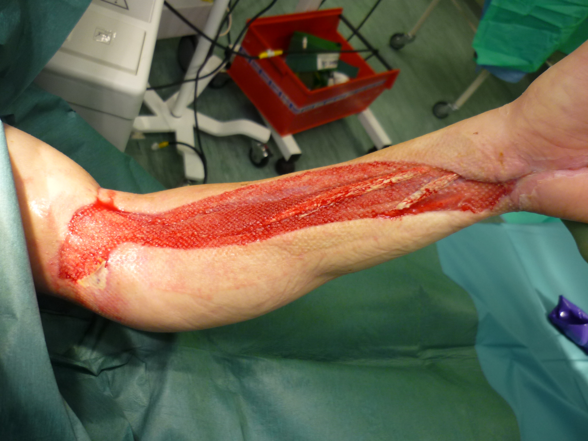
After flow returns, the ischemic muscle swells. Inside compartments closed by the fascia, this swelling raises pressure until it compromises capillary circulation, and the muscle that survived ischemia can die from reperfusion. The South African series indicates fasciotomy when there is distal swelling, ischemia for more than 4 to 6 hours or venous ligation, and recalls that compartment syndrome is a clinical diagnosis (Krüger et al., 2013).
In a Swedish series of 162 patients with vascular injuries from firearms, fasciotomy was needed in 17.9 in 100 patients, 29 of 162, and amputation in 1.2 in 100 (Nyberger et al., 2023). Fasciotomy leaves large wounds that need dressings and often skin grafting, but the cost of not doing it when indicated is much higher.
Compartment syndrome can appear even without arterial injury. A Turkish report describes compartment syndrome in the leg after arthroscopic removal of a low-velocity projectile from the knee, with an initially normal vascular examination confirmed by Doppler (Keskinbora et al., 2016). The example recalls that the limb must be reassessed after any procedure.
Clinical signs of compartment syndrome:
- Pain out of proportion to the apparent injury.
- Pain on passive stretching of the muscles of the compartment.
- Compartment tense on palpation.
- Paresthesia in the distribution of the nerves of the compartment.
- Loss of strength, at a later stage.
- Absence of pulse, a late sign and not required for the diagnosis.

Nerves, the damage that appears later
Nerve injuries accompany many vascular and bone injuries and often define limb function in the long term. In the series of distal humerus fractures, nerve injuries occurred in 52 in 100 patients and all were managed expectantly (Kauta et al., 2023).
A review of the timing of surgery in peripheral nerve injuries from firearms describes a spectrum that runs from direct laceration of the nerve to temporary neuropraxia, with complete spontaneous recovery, and concludes that the timing of intervention depends on clinical judgment, the mechanism of injury and a risk-benefit analysis in each patient (Tigre et al., 2024). Many injuries from the shock wave recover on their own; a nerve visibly severed during vascular surgery can be repaired or marked for later repair.
Distinguishing nerve injury from ischemia is part of the reasoning. Ischemia usually affects the entire distal segment, with a cold, pale foot, while nerve injury follows the distribution of a specific nerve, such as foot drop in fibular injury or claw hand in ulnar injury, with a warm, pink limb. When the deficit appears together with signs of ischemia, revascularization comes first, and the nerve is reassessed after flow returns.
Nerve recovery is slow and is followed with repeated clinical examination and, when necessary, electroneuromyography. Physical therapy, orthoses and control of neuropathic pain are part of treatment from the start, because joint stiffness and atrophy can limit function even when the nerve recovers.
The neurological examination on arrival is also important for practical reasons: a deficit present before any procedure will not later be attributed to the surgery, and it serves as a baseline for following recovery.
Outcomes and amputation
With rapid repair and an experienced team, most limbs are saved. In the Swedish series, amputation occurred in 1.2 per 100 patients (Nyberger et al., 2023). In the Somali series, with major delays in arrival, mortality was 5.8 per 100 and secondary amputation was 7.7 per 100, and the authors argue for revascularizing every viable limb (Hussein and Ali, 2024).
The factors that weigh most against the limb, according to the series cited, are ischemia time, combined injury of artery, vein, bone and nerve, extensive soft tissue loss and patient instability. These factors enter into the rare decision to amputate at the first surgery, made jointly by the vascular surgeon, the orthopedic surgeon and, when possible, with the patient and family.
The popliteal artery is the most difficult. In the South African series, 4 of 25 popliteal injuries required immediate amputation. When these injuries began to be operated on by vascular surgeons, reexploration fell from 34 to 10 per 100 and amputation from 16 to 11 per 100, a difference that was not statistically significant (Krüger et al., 2013).
Not every ischemia means a ruptured artery. One report describes a man with a gunshot wound to the thigh and acute ischemia from vasospasm of the superficial femoral artery, with CT angiography showing no flow in the middle segment. Flow returned quickly, the ischemia was treated without surgery, with serial examinations and Doppler, and he was discharged the next day (Goerlich et al., 2019). It is an exception, and it can only be accepted with close reassessment.
Antibiotic prophylaxis is also part of care, especially when there is a fracture. A prospective protocol with a single intravenous antibiotic dose for low-energy wounds to the limbs had deep infection requiring debridement in 0.8 per 100 patients and infections of all types in 11.1 per 100. Additional antibiotics outside the protocol did not reduce infection further (Nguyen et al., 2022). These data apply to low-energy wounds and do not automatically apply to complex vascular injuries. The regimen and duration follow the service protocol.
Who operates on the popliteal artery
A study at a large trauma center in Johannesburg followed all patients with penetrating arterial injury to the limbs for 18 months and changed one point of the protocol: popliteal artery injuries began to be operated on by vascular surgeons, while those of the axillary, brachial and femoral arteries remained with the trauma team (Krüger et al., 2013).
There were 113 patients with 116 injuries: 10 axillary, 47 brachial, 34 femoral and 25 popliteal (Krüger et al., 2013). The results in the axillary, brachial and femoral arteries were considered excellent and similar to the service's previous experience. In the popliteal artery, the reintervention rate fell from 34 to 10 percent, with a statistically significant difference, and the amputation rate fell from 16 to 11 percent, without a statistically significant difference (Krüger et al., 2013).
The authors themselves treat the result with caution. They suggest a possible trend toward better outcomes when the popliteal artery is operated on by vascular surgeons, but state that multicenter studies with more patients will be needed to confirm the hypothesis (Krüger et al., 2013). It is a good example of how to read a number: a drop that looks large may not be statistically significant when the sample is small.
For the student, the clinical lesson is that the popliteal artery remains the limb injury with the highest risk of limb loss, even in centers with extensive experience. This explains the insistence on recognizing hard signs early, measuring the ankle-brachial index when the signs are mild, controlling ischemia time and reassessing the compartments after revascularization. It also explains why many services call in vascular surgery right on arrival when the trajectory passes behind the knee.
How NEXO helps train this reasoning
Training with fictional clinical cases helps you recognize patterns before you encounter them on shift, as long as each clue is read calmly and compared with the hypotheses that are still open. In limb trauma, one clue may bring a weakening pulse, another a pain that worsens with stretching, and the combination points to a diagnosis that was not clear at the start. The discipline of reviewing the case before closing is covered in the text on diagnostic pause.
The texts on differential diagnosis with clues, diagnostic uncertainty and clinical decision-making also help. To understand points and rewards, see the NEXO Economy page. There are also tracks for those who research (Researcher), for those who review cases (Curator), for those who produce content (Creators) and for those who want to follow results (Closer).
Frequently asked questions
Does a palpable pulse rule out arterial injury?
No. Partial injuries, dissections and false aneurysms can maintain distal flow for a while. A pulse weaker than on the other side is a mild sign and calls for an ankle-brachial index or imaging.
Does every hematoma in the thigh need a CT angiography?
An expanding or pulsatile hematoma is a hard sign and indicates surgery. A small, stable hematoma is a mild sign; the decision to request imaging considers the trajectory, the index and the repeated examination.
Why is the popliteal artery so feared?
Because it sits trapped behind the knee, has few collaterals and divides just below into smaller branches. An occlusion there leaves the leg with little alternative circulation, and delay in repair quickly leads to muscle necrosis. Case series show the highest rates of compartment syndrome and amputation in this artery.
When should fasciotomy be performed?
When there are clinical signs of compartment syndrome, and preventively in high-risk situations, such as prolonged ischemia, combined artery and vein injury or venous ligation, according to the team's judgment.
Does the projectile need to be removed from the limb?
Most of the time, no. Removal is considered when the projectile is inside a joint, compresses a nerve, lies inside a vessel or causes symptoms.
Does an injured vein need to be repaired?
Large veins, such as the femoral and popliteal, are repaired when the patient is stable, to reduce edema and the risk of compartment syndrome. In the unstable patient, ligation may be necessary, and fasciotomy is then considered.
What is the difference between a false aneurysm and an arteriovenous fistula?
In a false aneurysm, blood leaves the artery and is contained by the surrounding tissues, forming a pulsatile sac. In an arteriovenous fistula, the injured artery and vein communicate directly, and arterial blood enters the vein, which can cause a thrill, a bruit and, over time, cardiac overload. Both can appear days or weeks after the injury.
What should be monitored after discharge?
Pulses, color and temperature of the limb, strength and sensation, wound healing and signs of infection. A pulsatile mass, a new bruit or pain when walking deserve early return and new imaging.
Take-home messages
- Control the bleeding before any examination.
- Look for hard signs and take the patient straight to surgery when present.
- Measure the ankle-brachial index when there are mild signs.
- Request CT angiography or Doppler according to the service and the clinical picture.
- Use a temporary shunt when an unstable fracture and arterial injury coexist.
- Examine and document nerve function on arrival.
- Reassess the compartments after revascularization.
If you like reasoning from clues, the case of the day is waiting on the NEXO home page.
References
- Goerlich CE, Challa AB, Malas MM. Acute limb ischemia from gunshot wound secondary to arterial vasospasm. J Vasc Surg Cases Innov Tech. 2019. PMID 31193453.
- Gopireddy DR, Kee-Sampson JW, Vulasala SSR, et al. Imaging of penetrating vascular trauma of the body and extremities secondary to ballistic and stab wounds. J Clin Imaging Sci. 2023. PMID 36751564.
- Hussein AM, Ali AA, Ahmed SA, et al. Our experience with blast and gunshot induced traumatic vascular injuries at Somalia's major vascular referral center. Sci Rep. 2024. PMID 38844514.
- Kauta N, Bott A, Majirija ET, et al. Surgical management of distal humerus gunshot fractures: descriptive case series. Eur J Orthop Surg Traumatol. 2023. PMID 37322263.
- Keskinbora M, Yalçin S, Oltulu İ, et al. Compartment Syndrome Following Arthroscopic Removal of a Bullet in the Knee Joint after a Low-Velocity Gunshot Injury. Clin Orthop Surg. 2016. PMID 26929809.
- Krüger A, Florido C, Braunisch A, et al. Penetrating arterial trauma to the limbs: outcome of a modified protocol. World J Emerg Surg. 2013. PMID 24305415.
- Montorfano MA, Pla F, Vera L, et al. Point-of-care ultrasound and Doppler ultrasound evaluation of vascular injuries in penetrating and blunt trauma. Crit Ultrasound J. 2017. PMID 28211004.
- Nguyen MP, Savakus JC, Simske NM, et al. Single dose IV Antibiotic for Low-Energy Extremity Gunshot Wounds: A Prospective Protocol. Ann Surg Open. 2022. PMID 37600115.
- Nyberger K, Caragounis EC, Djerf P, et al. Management and outcomes of firearm-related vascular injuries. Scand J Trauma Resusc Emerg Med. 2023. PMID 37420263.
- Stevenson T, Carr DJ, Gibb IE, et al. Preliminary effect of projectile yaw on extremity gunshot wounding in a cadaveric animal model: a serendipitous study. Int J Legal Med. 2020. PMID 32162008.
- Tigre JY, Puerto A, Khalafallah AM, et al. Timing of surgical intervention in peripheral nerve injuries from gunshot wounds: Management and review of the literature. Surg Neurol Int. 2024. PMID 38840615.