Skull gunshot wound: CT, prognosis, when to operate
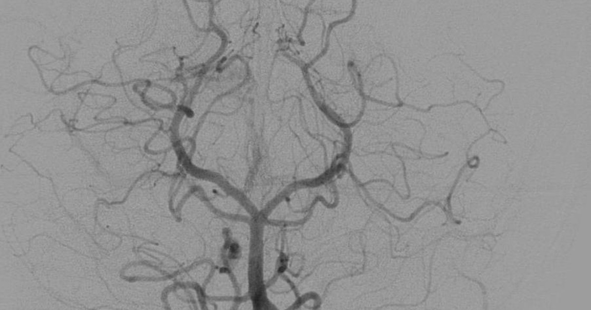
Table of contents
- The scale of the problem
- What happens inside the skull
- Initial assessment and neurological examination
- What to look for on the CT scan
- Prognostic factors
- When surgery tends to help
- Infection, seizures and other complications
- Limits of care and organ donation
- How NEXO helps train this reasoning
- Frequently asked questions
- Take-home messages
- References
The following scene is fictional and was written for teaching. A 27-year-old man arrives at the emergency department brought by family members, with a gunshot wound to the right temporal region. He opens his eyes to pain, makes incomprehensible sounds and localizes the stimulus with his left hand. The pupils are isocoric and react to light. Blood pressure is 150 over 80 mmHg and heart rate is 58 beats per minute. The CT scan shows a track that enters through the temporal bone, crosses the temporal lobe and stops before the midline, with a small hematoma and bone fragments along the track. The question that hangs in the room is always the same: does this patient have a chance?
This text summarizes what the literature shows about gunshot wounds to the head: how to interpret the CT scan, which factors weigh on prognosis, when surgery tends to help and which complications appear afterwards. The content is educational, aimed at students and health professionals, and does not replace local protocols or the assessment of the neurosurgery team.
Key points
- About 90 out of every 100 victims of gunshot wounds to the skull die before reaching the hospital, and about half of those who arrive die in the emergency department.
- A low Glasgow Coma Scale score after resuscitation, fixed pupils, a transventricular or bihemispheric track and injury to deep structures indicate a worse prognosis.
- Patients with a Glasgow score of 8 or more usually receive aggressive treatment; with 3 to 5 and signs of diffuse injury, the decision is individualized.
- The current trend is less aggressive debridement of deep fragments and more consistent antibiotic prophylaxis.
- Vascular injury should be sought when the track passes near large vessels or crosses hemispheres.
- Organ donation is a legitimate goal when the injury is not survivable.
The scale of the problem
Gunshot wounds to the head are the most lethal form of traumatic brain injury. A review on the management of these injuries estimates that about 90 out of every 100 victims die before reaching the hospital and that approximately half of those who arrive die in the emergency department (Alvis-Miranda et al., 2015). Those who reach the hospital alive are therefore already a selected group, and it is in this group that clinical decisions make a difference.
A national analysis of gunshot wounds to the head and neck, based on United States hospitalization data, found in-hospital mortality of 25.7 per 100 patients with these injuries, compared with 2.7 per 100 among patients with gunshot wounds to other regions (Menezes et al., 2023). The difference shows the specific weight of the skull among projectile wounds.
In the Helsinki series, which followed gunshot wounds to the brain over several years, 64 patients reached the hospital alive. Mortality among those operated on was 32 per 100, while among those who received no active treatment it approached 100 per 100 (Frösen et al., 2019). The authors observed that most wounds were self-inflicted, with a contact shot from a handgun, a pattern that changes the injury profile and the prognosis.
What happens inside the skull
The brain is a poorly elastic tissue held inside a rigid box. When a projectile crosses the skull, it produces a permanent cavity, the track of destroyed tissue, and a temporary cavity, a rapid expansion that compresses and stretches the surrounding tissue. A review on the physics of traumatic brain injury describes that, in low-velocity injuries, the damage stays localized along the track, while in high-velocity injuries cavitation is added, which does not accompany low-velocity injuries (Young et al., 2015).
Beyond the direct injury, there is a cascade of secondary events: edema, intraparenchymal and extra-axial hematomas, intraventricular hemorrhage, intracranial hypertension and coagulation changes. Bone fragments driven into the brain act as secondary projectiles and are a source of infection. The path may cross the paranasal and mastoid sinuses, opening the way for cerebrospinal fluid leak and meningitis.
The mechanisms of secondary injury that are of most concern in the first hours are:
- Progressive cerebral edema, which raises the pressure inside the skull.
- Expansion of hematomas, mainly in patients with coagulopathy.
- Hypotension and hypoxia, which reduce the oxygen supply to an already injured brain.
- Acute hydrocephalus from blood in the ventricles.
- Seizures, which increase oxygen consumption and intracranial pressure.
- Early infection from the material carried into the path.
Much of the care in the first hours consists of preventing these complications. The primary injury has already happened at the moment of the shot; what the team can change is what comes after.
For this reason, severity does not depend only on the entry point. A projectile that crosses only one lobe, without crossing the midline or reaching the ventricles, has very different consequences from one that crosses both hemispheres or passes through the brainstem. This concept guides both the reading of the image and the conversation with the family.


Initial assessment and neurological examination
The assessment begins with the usual trauma sequence: airway, breathing, circulation, neurological status and exposure. Patients with a significant drop in level of consciousness usually need a definitive airway, and hypotension and hypoxia should be avoided because they worsen secondary injury. Scalp bleeding can be heavy and needs rapid control.
The neurological examination should be recorded after correction of hypotension, hypoxia and the effect of sedatives, because the Glasgow coma scale obtained after resuscitation better predicts the extent of the initial injury (Qi and Li, 2021). The elements that weigh most are:
- Glasgow coma scale score after resuscitation.
- Pupil size and reactivity, including asymmetry.
- Signs of intracranial hypertension, such as bradycardia with high blood pressure.
- Focal deficits, which help locate the injury.
- Leakage of cerebrospinal fluid or brain tissue through the wound, the nose or the ear.
- Coagulopathy, which should be sought and corrected early.
In children, the assessment follows the same logic, but the coma scale needs adaptation for age, and the volume of blood lost from a scalp wound weighs proportionally more. In older adults, the use of anticoagulants and antiplatelet agents is frequent and increases the risk of hematoma expansion, so the medication in use should be checked as soon as they arrive, along with coagulation tests.
In the fictional opening scene, bradycardia with high blood pressure deserves immediate attention. It is a possible sign of intracranial hypertension and may require rapid measures while the neurosurgery team assesses. The text on diagnostic pause discusses how to keep a single reassuring finding, such as normal pupils, from hiding another alarming one.
What to look for on the CT scan
Before getting to the image, a practical reminder is worthwhile: plain skull radiography can locate the projectile, but it does not show the brain injury. It has little role when CT is available and should not delay it.
Non-contrast head CT is the initial exam. It shows the path, the location of the projectile and fragments, hematomas, edema, midline shift and ventricular involvement. A review of cases and literature highlights that the assessment of most of these wounds requires very rapid imaging on arrival, followed by active treatment against infection, seizures and intracranial hypertension (Qi and Li, 2021).
Some findings deserve attention when reading:
- A path that crosses the midline or passes through both hemispheres.
- Passage through the ventricles or intraventricular hemorrhage.
- Involvement of deep structures, such as the basal ganglia, thalamus and brainstem.
- Multilobar injury.
- Hematoma with mass effect, which may be drained.
- A path near the skull base, the cavernous sinus or the venous sinuses.
Suspected vascular injury calls for CT angiography or angiography. The review by Kazim and colleagues recommends cerebral angiography when there is high suspicion of vascular injury (Kazim and colleagues, 2011). A review of craniocervical vascular imaging lists bihemispheric trajectory among the risk factors and describes that, among the vascular injuries found, arteriovenous fistula accounted for 86 in 100 and pseudoaneurysm for 12 in 100 (Kee-Sampson and colleagues, 2022). Traumatic pseudoaneurysm can rupture days later, so it must not be forgotten.


Prognostic factors
Few topics in trauma generate as much discussion as the prognosis of gunshot wounds to the head. The literature converges on some factors. The review by Kazim and colleagues points to a Glasgow Coma Scale of 3 with fixed and dilated pupils, self-inflicted wound, coagulopathy and high intracranial pressure as markers of poor prognosis (Kazim and colleagues, 2011). The review by Alvis-Miranda and colleagues adds multilobar injury and intraventricular hemorrhage (Alvis-Miranda and colleagues, 2015).
In the Helsinki series, the factors associated with mortality were a Glasgow score of 8 or less on admission, transventricular trajectory and injury to deep structures. The authors concluded that a contact gunshot wound from a short firearm has high mortality, but can survive with a reasonable outcome when the injury is restricted to one lobe, without significant damage to deep structures or the brainstem (Frösen and colleagues, 2019).
| Factor | Tends to favor | Tends to worsen |
|---|---|---|
| Glasgow after resuscitation | 8 or more | 3 to 5 |
| Pupils | Reactive and symmetric | Fixed, bilaterally dilated |
| Trajectory | Restricted to one lobe | Bihemispheric, transventricular, multilobar |
| Deep structures | Spared | Basal ganglia, thalamus or brainstem affected |
| Intraventricular blood | Absent | Present |
| Coagulation | Normal | Coagulopathy |
| Context | Wound not self-inflicted | Self-inflicted wound |
These factors are not sentences. A report of four patients with a Glasgow score below 5 on admission, all submitted to decompressive hemicraniectomy, showed that three were functionally independent one year later (Lin and colleagues, 2012). The authors concluded that victims of these wounds can have good outcomes despite a very low Glasgow score on arrival. Selected reports do not define management, but they remind us that prognosis is probability, not certainty. The text on diagnostic uncertainty explores this idea further.
When surgery tends to help
Before talking about surgery, it is worth remembering that medical treatment of intracranial hypertension is a central part of care. Head of bed at about 30 degrees, head aligned, adequate sedation and analgesia, control of temperature, blood glucose and sodium, ventilation that avoids both hypoxia and prolonged hyperventilation, and correction of coagulopathy are measures that are usually started early. Intracranial pressure monitoring may be indicated according to the examination and imaging. These measures do not repair the primary injury, but they buy time and reduce secondary injury.
The most accepted surgical indications include hematoma with mass effect, refractory intracranial hypertension with an indication for decompression, accessible fragments with risk of infection, a wound with exposure of brain tissue and a cerebrospinal fluid leak that does not resolve. The review by Alvis-Miranda and colleagues notes that patients with a Glasgow score of 3 to 5 are often not treated aggressively, while patients with 8 or more usually receive aggressive treatment (Alvis-Miranda and colleagues, 2015). Between these extremes, the decision depends on the set of findings.
The extent of debridement has changed over the decades. The review by Kazim and colleagues describes the current trend toward less aggressive debridement of deep bone and metal fragments, combined with more intense antibiotic prophylaxis (Kazim and colleagues, 2011). Searching for every deep fragment can cause more injury than the fragment itself.
When removal of the projectile is indicated, technique matters. In a prospective cohort of 28 patients with gunshot wounds to the head, the authors compared frame-based and frameless stereotaxy, fluoroscopy-guided surgery and freehand surgery. Planned stereotaxy failed to locate the projectile in three of eight patients, who required conventional craniotomy, and neuronavigation had an excellent result in five of nine (Elserry et al., 2013). The authors discuss that the choice depends on location and available resources.


Infection, seizures and other complications
Infection is a constant threat, because the projectile carries hair, skin, bone and clothing fragments into the brain. Tracks that pass through the paranasal sinuses or the mastoid increase the risk of cerebrospinal fluid leak and meningitis. The review by Kazim et al. cites a historical series with cerebrospinal fluid leak in 28 of every 100 patients (Kazim et al., 2011).
Post-traumatic epilepsy is also frequent. The same review states that the risk of epilepsy after penetrating brain injury is high and that prophylactic anticonvulsant use is recommended (Kazim et al., 2011). The choice of drug and duration follow local protocols and the neurological assessment.
There are also late complications that surprise. One report describes tension pneumocephalus about 16 weeks after bifrontal craniectomy for a self-inflicted injury. The patient had a month of rhinorrhea, postnasal drip and cough before feeling a sensation of expansion in the forehead (Wang et al., 2016). The authors note that tension pneumocephalus does not always appear soon after trauma or surgery and can develop insidiously.
Those who survive usually need prolonged rehabilitation. Motor deficits, language changes, memory and attention disorders, mood changes and late epilepsy are frequent and change the lives of the patient and the family. Outpatient follow-up with neurology, neurosurgery, physical therapy, speech therapy, occupational therapy and mental health is part of treatment, not an accessory. When the injury was self-inflicted, mental health care and prevention of a new episode of self-harm must be planned from admission onward.
Other complications described include brain abscess, hydrocephalus, migration of the projectile within the parenchyma or ventricles, and lead poisoning when the projectile stays in contact with the cerebrospinal fluid. Clinical and imaging follow-up is part of treatment.
Limits of care and organ donation
Before any conversation about limits, the team must be sure it has completed the full assessment. This includes correcting hypothermia, hypotension, hypoxia and the effect of sedatives, reviewing the imaging with neurosurgery and, when in doubt, allowing time for the examination to become clear. Hasty decisions in the first hours, based only on the appearance of the wound or the presumed severity of the mechanism, can take away the chance of patients who would have evolved better than expected.
Not every gunshot wound to the head is survivable, and recognizing this is part of good care. In a series of patients with gunshot wounds to the brain, only 2.1 of every 100 of those who required cardiopulmonary resuscitation survived (Robinson et al., 2019). The same study showed that the organ donation rate was 10.3 of every 100 overall and 58.9 of every 100 among eligible patients.
The review by Alvis-Miranda et al. recognizes organ donation as a legitimate goal in the management of patients with non-survivable injuries (Alvis-Miranda et al., 2015). This means maintaining hemodynamic stability and oxygenation while the family is heard and the brain death and donation protocols are followed.
The conversation with the family must be honest about uncertainty. Phrases like "there is nothing to do" are inaccurate and hurt. It is more correct to explain what the imaging and the exam show, what the probabilities are and what care is still being offered. For those who want to train how to reason in the face of incomplete data, the text on clinical decision-making is a good complement.
Antibiotic, intracranial pressure and late vascular injury
The review by Kazim and colleagues brings together three points of post-surgery care that usually end up in the background. The first is infection. In the military series from the era before antibiotics, infectious complications reached 58.8 percent of the wounded, and in a large civilian review with broad-spectrum antibiotics the rate was between 1 and 5 percent (Kazim and colleagues, 2011). That is why the authors recommend starting a broad-spectrum antibiotic in every penetrating brain injury, as early as possible, with cephalosporins as the most cited option; the exact regimen follows the local protocol (Kazim and colleagues, 2011).
The second point is intracranial pressure. In severe closed brain injury, monitoring is a well-documented predictor of prognosis, and control of intracranial hypertension is associated with better recovery. In penetrating injury, however, clinical data on the indication and use of monitoring are scarce, and the published studies come mostly from military settings (Kazim and colleagues, 2011). In practice, the decision is usually extrapolated from closed trauma and individualized.
The third point is the vascular injury that appears late. According to the same review, some injuries appear weeks or months after the trauma, and a normal initial angiogram does not close the question. When suspicion persists, repeating the exam two to three weeks later is recommended (Kazim and colleagues, 2011). Traumatic intracranial aneurysm is the most described vascular injury after penetrating injury, and about 20 percent of traumatic aneurysms after brain injury are caused by a penetrating mechanism (Kazim and colleagues, 2011).
For those who study, the lesson is that the reasoning does not end at the CT scan on arrival. Fever, neurological worsening or new bleeding days later call for reopening the hypotheses of infection, intracranial pressure and late vascular injury.
How NEXO helps train this reasoning
Training with fictional clinical cases helps recognize patterns before meeting them on shift, as long as each clue is read calmly and compared with the hypotheses that are still open. In cases of head trauma, the clues usually combine vital signs, neurological exam and imaging findings, and the challenge is to put these pieces together without sticking to the first impression. It is the same training described in the text on differential diagnosis with clues.
The text on blast trauma complements this one, because explosive fragments also cause penetrating head injuries. To understand how points and rewards work, see the NEXO Economy page. There are also tracks for those who research (Researcher), for those who review cases (Curator), for those who produce content (Creators) and for those who want to follow results (Closer).
Frequently asked questions
Is every gunshot wound to the skull fatal?
No. Mortality is very high, especially before arrival at the hospital, but there are survivors, including with good functional recovery. Injuries restricted to one lobe, without passing through the ventricles or deep structures, have a better prognosis.
Does the projectile always need to be removed from the brain?
No. The current tendency is to avoid aggressive debridement of deep fragments. Removal is considered when the projectile is accessible, migrates, causes mass effect, is associated with infection or is in contact with the cerebrospinal fluid.
Why should the Glasgow scale be measured after resuscitation?
Because hypotension, hypoxia, intoxication and sedatives lower the level of consciousness for reasons that do not depend on the brain injury. The score after correcting these causes reflects the severity of the injury better.
When should vascular injury be considered?
When the track passes near major arteries, the skull base or the venous sinuses, crosses the hemispheres, or when there is unexplained bleeding or late deterioration. CT angiography or angiography can identify pseudoaneurysms and fistulas.
Why talk about organ donation in this context?
Because some of the patients who arrive at the hospital have non-survivable injuries and may progress to brain death. Maintaining support while the family is heard allows the patient's wishes to be respected and donation to be offered when it is possible.
What changes in pre-hospital care?
The focus is on maintaining an adequate airway, oxygenation and blood pressure, controlling scalp bleeding and transporting the patient quickly to a center with neurosurgery. Objects or projectiles stuck in the wound should not be removed on site. Recording the level of consciousness and the pupils at the scene helps the team receiving the patient a great deal.
Do normal pupils rule out severe injury?
No. Reactive pupils are a good sign, but the patient can deteriorate in the following hours due to hematoma expansion or edema. Bradycardia with high blood pressure, a worsening level of consciousness and new deficits call for immediate reassessment and, often, a new CT scan.
Take-home messages
- Correct hypotension and hypoxia before recording the Glasgow scale.
- Assess pupils, signs of intracranial hypertension and coagulation.
- Read the track, the ventricles, the deep structures and the hematomas on the CT scan.
- Consider CT angiography when the track passes near major vessels or crosses the hemispheres.
- Remember infection and seizure prophylaxis according to local protocol.
- Watch for late complications, such as cerebrospinal fluid leak, abscess and pneumocephalus.
- Talk to the family honestly and consider donation when the injury is not survivable.
If you like reasoning from clues, the case of the day is waiting on the NEXO home page.
References
- Alvis-Miranda HR, Adie Villafañe R, Rojas A, et al. Management of Craniocerebral Gunshot Injuries: A Review. Korean J Neurotrauma. 2015. PMID 27169063.
- Elserry T, Anwer H, Esene IN. Image guided surgery in the management of craniocerebral gunshot injuries. Surg Neurol Int. 2013. PMID 24349869.
- Frösen J, Frisk O, Raj R, et al. Outcome and rational management of civilian gunshot injuries to the brain-retrospective analysis of patients treated at the Helsinki University Hospital from 2000 to 2012. Acta Neurochir (Wien). 2019. PMID 31129782.
- Kazim SF, Shamim MS, Tahir MZ, et al. Management of penetrating brain injury. J Emerg Trauma Shock. 2011. PMID 21887033.
- Kee-Sampson JW, Gopireddy DR, Vulasala SSR, et al. Role of imaging in penetrating vascular injuries of the craniocervical region. J Clin Imaging Sci. 2022. PMID 36601604.
- Lin DJ, Lam FC, Siracuse JJ, et al. "Time is brain" the Gifford factor - or: Why do some civilian gunshot wounds to the head do unexpectedly well? A case series with outcomes analysis and a management guide. Surg Neurol Int. 2012. PMID 23061014.
- Menezes JM, Batra K, Zhitny VP. A Nationwide Analysis of Gunshot Wounds of the Head and Neck: Morbidity, Mortality, and Cost. J Craniofac Surg. 2023. PMID 36927798.
- Qi H, Li K. Civilian gunshot wounds to the head: a case report, clinical management, and literature review. Chin Neurosurg J. 2021. PMID 33531086.
- Robinson LA, Turco LM, Robinson B, et al. Outcomes in patients with gunshot wounds to the brain. Trauma Surg Acute Care Open. 2019. PMID 31799416.
- Wang A, Solli E, Carberry N, et al. Delayed Tension Pneumocephalus following Gunshot Wound to the Head: A Case Report and Review of the Literature. Case Rep Surg. 2016. PMID 28074167.
- Young L, Rule GT, Bocchieri RT, et al. When physics meets biology: low and high-velocity penetration, blunt impact, and blast injuries to the brain. Front Neurol. 2015. PMID 25999910.