Digital health and AIEndocrinologyPrimary care
Autonomous AI in diabetic retinopathy screening
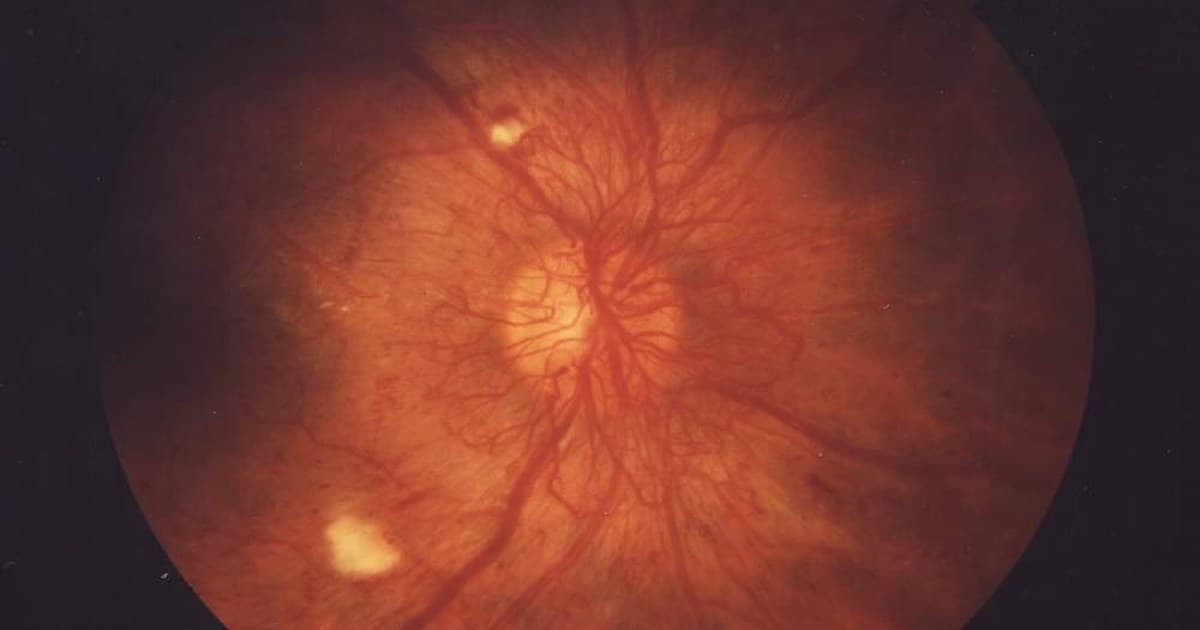



Table of contents
- Why screen for diabetic retinopathy
- What the WHO expects from a screening program
- From algorithm to autonomous diagnosis
- Sensitivity and specificity in screening
- Performance outside the original study
- What happens with referral
- Limits and precautions
- How NEXO helps train this reasoning
- Frequently asked questions
- Take-home messages
- References
In a fictional case, a 52-year-old man with type 2 diabetes for six years goes to the primary care unit to renew prescriptions. He sees well, has no visual complaint and has not had a fundus exam in three years, because the nearest ophthalmologist is a two-hour trip away. In the room next to the office there is a retinal camera. A technician from the unit itself photographs both eyes, a program analyzes the images and, a few minutes later, the result appears in the medical record: more than mild diabetic retinopathy detected, refer to the specialist.
The result came out before any doctor looked at the photos. This is what is called autonomous diagnosis by artificial intelligence (AI): the system decides on its own whether the exam is positive or negative, and the person operating the camera does not need to know how to interpret the retina. The World Health Organization (WHO) lists eye examination to screen for retinopathy, which causes blindness, among the additional care that a person with diabetes may need (WHO, Diabetes).
This text uses the WHO as its axis and goes through three questions: how the first autonomous diagnosis came about, what sensitivity and specificity mean when the goal is screening, and what happens to the patient after the system says to refer.
Key points
- In 2016, a deep learning algorithm detected referable diabetic retinopathy in photographs with 97.5 percent sensitivity and 93.4 percent specificity at a cutoff chosen for screening, in a retrospective study (Gulshan et al., 2016).
- The pivotal 2018 study, at ten primary care sites in the United States, showed 87.2 percent sensitivity and 90.7 percent specificity, and led to the authorization of the first autonomous diagnosis by AI (Abràmoff et al., 2018).
- In a randomized trial with young people aged 8 to 21, autonomous examination at the site itself raised completion of the eye exam within six months from 22 to 100 percent (Wolf et al., 2024).
- For the WHO, screening requires a complete pathway, with referral, confirmation and treatment, and not just a test (WHO Europe, diabetic retinopathy screening guide, 2020).
- Why screen for diabetic retinopathy
- What the WHO expects from a screening program
- From algorithm to autonomous diagnosis
- Sensitivity and specificity in screening
- Performance outside the original study
- What happens with referral
- Limits and precautions
- How NEXO helps train this reasoning
- Frequently asked questions
- Take-home messages
Why screen for diabetic retinopathy
The number of people living with diabetes went from more than 200 million in 1990 to 830 million in 2022, and the increase was faster in low- and middle-income countries (WHO, Diabetes). In 2022, 14 percent of adults aged 18 or older were living with diabetes (WHO, Diabetes).
Diabetic retinopathy is among the leading causes of visual impairment and blindness in the world, alongside refractive errors, cataract, glaucoma and age-related macular degeneration (WHO, Blindness and vision impairment). Among the billion people whose visual impairment could have been prevented or has not yet been treated, the WHO attributes 3.9 million to diabetic retinopathy (WHO, Blindness and vision impairment).
The central point for screening is that the disease is usually silent at the start. The WHO highlights that, in conditions such as diabetic retinopathy, early detection and timely treatment are decisive for avoiding irreversible vision loss (WHO, Blindness and vision impairment). The WHO European guide defines the goal of screening as reducing the risk of visual impairment and blindness in people with diabetes without visual symptoms, through the identification and treatment of sight-threatening retinopathy (WHO Europe, diabetic retinopathy screening guide, 2020).
Control of blood glucose, blood pressure and lipids remains part of care, and what changed in the general diabetes recommendations is summarized in ADA 2026: what changed in diabetes. Here the focus is different: who looks at the retina, and how.
The specialist bottleneck
The traditional exam depends on someone trained to examine or photograph the fundus and interpret the finding. The WHO guide cites a systematic review in which the sensitivity of direct ophthalmoscopy performed by family physicians ranged from 25 to 66 percent, versus 43 to 79 percent when performed by ophthalmologists (WHO Europe, diabetic retinopathy screening guide, 2020). When resources allow, the same guide points to digital retinal photography as the preferred method, because it produces quality images that can be stored and audited.
Adherence to the exam is also low. Leong and colleagues report that, in the United States, adherence to eye exam recommendations in people with diabetes is as low as 20 percent in some studies (Leong et al., 2026).
What the WHO expects from a screening program
The 2020 European guide applies the ten Wilson and Jungner screening principles, published by the WHO in 1968, to diabetic retinopathy (WHO Europe, diabetic retinopathy screening guide, 2020).
The guide states that effective screening requires a pathway, not just a test (WHO Europe, diabetic retinopathy screening guide, 2020). This pathway runs from identifying the population to invitation, testing, referral of positives, diagnostic confirmation, treatment and recording of outcomes. The WHO general guide to screening programs repeats the logic: positives are referred, false positives are identified at the diagnostic stage and outcomes are analyzed to recognize false negatives (WHO Europe, screening programs guide, 2020).
Referral threshold and interval
The degree of retinopathy from which the patient is referred is called the referral threshold, and it can vary between countries (WHO Europe, diabetic retinopathy screening guide, 2020). A low threshold, such as mild non-proliferative retinopathy, can overload services; a high threshold, such as severe non-proliferative retinopathy, risks delaying the detection of advanced disease.
As for the interval, most of the studies cited by the WHO conclude that screening should be repeated every one or two years (WHO Europe, diabetic retinopathy screening guide, 2020).
Coverage as an indicator
The England program screened 2,847,149 people with diabetes in 2018 and 2019, with coverage of 83 percent, according to the guide (WHO Europe, diabetic retinopathy screening guide, 2020). The same document reports that, after seven years of screening, diabetic retinopathy ceased to be the most common cause of blindness in the English working-age population.
From algorithm to autonomous diagnosis
The 2016 study
The work by Gulshan and colleagues, published in 2016, trained a neural network with 128,175 retinal photographs graded three to seven times by 54 ophthalmologists and final-year residents (Gulshan et al., 2016). The target was referable diabetic retinopathy, defined as moderate or worse retinopathy, referable macular edema, or both. Validation used two sets: 9,963 images from 4,997 patients at screening services in the United States and 1,748 images from 874 patients at three hospitals in France.
The algorithm was evaluated at two cut-off points. At the point chosen for high specificity, sensitivity was 90.3 percent and specificity 98.1 percent in the first set (Gulshan et al., 2016). At the point chosen for high sensitivity, sensitivity rose to 97.5 percent and specificity fell to 93.4 percent (Gulshan et al., 2016). The authors explain that they chose the second point, with sensitivity of 97 percent in the development set, because high sensitivity is a prerequisite for a screening tool (Gulshan et al., 2016).
The study was retrospective, and the reference was the decision of the majority of ophthalmologists who read the images. The authors conclude that it remained to be seen whether the algorithm would work in clinical practice and whether it would improve care (Gulshan et al., 2016).
The pivotal 2018 study
The prospective answer came with the study by Abràmoff and colleagues. Between January and July 2017, 900 people with diabetes and no history of retinopathy were enrolled at ten primary care sites in the United States (Abràmoff et al., 2018). The camera was operated by the staff already at the sites, after standardized training, and the reference was demanding: stereoscopic wide-field photographs and optical coherence tomography of the macula, taken by certified photographers and graded by an independent reading center.
The target was more than mild retinopathy, defined as level 35 or higher on the ETDRS scale, macular edema, or both, in at least one eye (Abràmoff et al., 2018). Of the 900 participants, 819 could be fully analyzed, and 198 of them, or 23.8 percent, had that degree of disease (Abràmoff et al., 2018).
The system exceeded the three goals set before the study: sensitivity of 87.2 percent, above the 85 percent goal; specificity of 90.7 percent, above the 82.5 percent goal; and a rate of gradable images of 96.1 percent (Abràmoff et al., 2018). To obtain a result, 23.6 percent of participants needed pupil dilation (Abràmoff et al., 2018). Based on these data, the United States regulatory agency authorized use of the system by health professionals, making it the first autonomous AI diagnostic system authorized in any area of medicine.
Sensitivity and specificity in screening
The WHO guide defines sensitivity as the ability of the test to detect the disease when it is present, and specificity as the ability to indicate that nothing is wrong when the disease is absent (WHO Europe, diabetic retinopathy screening guide, 2020).
In screening, the two errors have different consequences. The WHO guide to screening programs notes that results from healthy and sick people always overlap, so some healthy people receive a positive result, the false positives, and some sick people receive a negative result, the false negatives (WHO Europe, screening programs guide, 2020). A false negative sends someone who needed treatment back to the routine interval; a false positive takes a specialist slot and causes anxiety.
The same algorithm, two cutoffs
The 2016 study shows that sensitivity and specificity are not fixed properties of the algorithm. By changing the cutoff, the same model went from 90.3 percent sensitivity with 98.1 percent specificity to 97.5 percent sensitivity with 93.4 percent specificity (Gulshan et al., 2016). Whoever implements the system chooses where to sit on that trade-off, and screening usually favors sensitivity.
Prevalence and predictive value
Prevalence changes how the result is read. With about 8 percent referable retinopathy in the images, the Gulshan algorithm had a negative predictive value of 99.8 percent in the American set (Gulshan et al., 2016). The reasoning is the same as for any screening test, in which the result must be read in light of the pre-test probability, as in the text on AUDIT, CAGE and ASSIST as pre-test probability.
An illustrative calculation helps. Applying the 87.2 percent sensitivity and 90.7 percent specificity from the pivotal study to the 23.8 percent prevalence observed in it (Abràmoff et al., 2018), of every thousand people screened about 238 would have the disease: approximately 208 would be detected and 30 would receive a negative result. Among the 762 without the disease, about 71 would be referred unnecessarily.
Images the system cannot read
There is a third result beyond positive and negative: the image of insufficient quality. In the pivotal study, among the 33 participants whose images the system considered insufficient, 30 percent had more than mild retinopathy, a proportion comparable to the rest of the sample (Abràmoff et al., 2018). This group cannot simply go home with advice to repeat the exam in a year.
Gulshan and colleagues evaluated the effect of counting the ungradable image as a reason for referral. In that scenario, at the high-sensitivity point, sensitivity was 96.7 percent and specificity fell to 84.0 percent (Gulshan et al., 2016). In the practice described by Leong and colleagues, referral was made both after a positive result and after a non-diagnostic result (Leong et al., 2026).
Comparison between the main performance studies
| Study | Design and location | Target | Main result |
|---|---|---|---|
| Gulshan 2016 | Retrospective; images from the United States and France | Referable retinopathy | Sensitivity of 97.5 and specificity of 93.4 percent at the screening point |
| Abràmoff 2018 | Prospective; 10 primary care services in the United States | More than mild retinopathy | Sensitivity of 87.2 and specificity of 90.7 percent; analyzable images in 96.1 percent |
| Ipp 2021 | Prospective; 15 centers in the United States, per eye | More than mild retinopathy | Sensitivity of 95.5 and specificity of 85.0 percent without dilation |
| Lee 2021 | Retrospective; seven algorithms on images from veterans | Referable retinopathy | Sensitivities from 50.98 to 85.90 percent across the algorithms |
The numbers in the table come from Gulshan et al., 2016, Abràmoff et al., 2018, Ipp et al., 2021 and Lee et al., 2021. The references, the populations and the units of analysis differ, so the table guides the reading but does not allow systems to be added up or ranked.
Performance outside the original study
The second pivotal study of an autonomous system, published by Ipp and colleagues, included 942 adults with diabetes at 15 centers in the United States, between April 2017 and May 2018 (Ipp et al., 2021). The centers were six primary care, six general ophthalmology and three retina. The analysis was done per eye, not per person, which prevents direct comparison of the numbers with those of the 2018 study.
With undilated pupils, sensitivity for more than mild retinopathy was 95.5 percent and specificity 85.0 percent; for vision-threatening retinopathy, 95.1 and 89.0 percent (Ipp et al., 2021). The system was able to grade 87.4 percent of eyes without dilation, and 97.4 percent when eyes with insufficient images were dilated (Ipp et al., 2021).
Seven algorithms, the same patients
A study by Lee and colleagues tested seven algorithms from five companies, one of them approved by the American agency, on 311,604 images from 23,724 veterans screened in two health systems in the United States between 2006 and 2018 (Lee et al., 2021).
Negative predictive values were high, from 82.72 to 93.69 percent, but sensitivities varied widely, from 50.98 to 85.90 percent (Lee et al., 2021). Most algorithms did not outperform human readers, and one of them had a sensitivity of 74.42 percent for proliferative retinopathy, lower than that of the service readers (Lee et al., 2021). The authors conclude that every algorithm should undergo rigorous testing with real-world data before entering clinical practice.
What the services that adopted them report
The review by Teng and colleagues gathered the literature and interviews with ophthalmologists who led the implementation in academic health systems (Teng et al., 2026). The services that adopted the systems reported gradable images without dilation in 49 to 75 percent of exams, sensitivity of 87 to 100 percent and specificity of 60 to 91 percent (Teng et al., 2026).
The spread is expected when the camera leaves a controlled protocol and enters a service with another team and another population, a problem also discussed in AI in nephrology and in AI in ultrasound.
What happens with referral
A positive result only protects vision if the patient reaches the specialist and is treated. The WHO guide calls for referral pathways defined with professional societies, for positives to be assessed quickly and for the time between referral and treatment to be agreed and monitored (WHO Europe, diabetic retinopathy screening guide, 2020).
The trial with young people
The ACCESS study by Wolf and colleagues randomized young people aged 8 to 21 with type 1 or type 2 diabetes at an academic pediatric diabetes center (Wolf et al., 2024). The intervention group had the autonomous AI exam at the visit itself; the control group received standardized guidance and a referral to an eye care professional.
The primary outcome was completion of the eye exam within six months: 100 percent in the intervention group, with 81 participants, versus 22 percent in the control group, with 83 (Wolf et al., 2024). In the intervention group, 25 participants had an abnormal result, and 16 of them, or 64 percent, saw an eye care professional within six months (Wolf et al., 2024). In the control group, 18 participants saw that professional, 22 percent of the group, and none had diabetic eye disease (Wolf et al., 2024).
The result shows both sides of referral. The AI closed the exam gap, but one in three young people with an abnormal result still had not reached the specialist within the study period (Wolf et al., 2024).
Who reaches the specialist
Leong and colleagues reviewed the records of 3,745 adults with diabetes referred from primary care in a Baltimore health system to a tertiary eye center between August 2020 and September 2022 (Leong et al., 2026). There were 3,352 patients through the usual referral and 393 through the AI pathway, in which referral occurred after a positive result, defined as level 35 or higher on the ETDRS scale or macular edema, or after a non-diagnostic result.
After propensity score weighting and adjustment for social and clinical factors, patients on the AI pathway were more likely to be African American, with an odds ratio of 1.15 and a 95 percent confidence interval between 1.02 and 1.29 (Leong et al., 2026). For public Medicaid insurance coverage, there was no significant difference. The authors classify the analysis as exploratory and state that it does not allow causal inference.
Referral is not the end
The points below summarize what the sources describe as necessary after a positive result:
- Having a referral pathway agreed with specialists and a monitored time frame between referral and treatment (WHO Europe, diabetic retinopathy screening guide, 2020).
- Treating a non-diagnostic result as a reason for referral or a repeat attempt, and not as a normal exam (Leong et al., 2026).
- Aligning the tool with the clinic workflow, making it easier to contact the patient and to make referrals, and training the team continuously (Teng et al., 2026).
- Also informing the patient and the physician of a negative result, and reviewing glycemic, blood pressure and lipid control when there is mild retinopathy below the threshold (WHO Europe, diabetic retinopathy screening guide, 2020).
When the target is not met and the approach stays the same, the problem is no longer the test but the system, a topic discussed in clinical inertia.
Limits and precautions
The articles themselves point out what autonomous diagnosis still does not solve. The algorithm was trained for retinopathy and macular edema, may miss other retinal diseases and does not replace a complete ophthalmological exam (Gulshan et al., 2016). In the 2018 pivotal study, all participants had no prior history of retinopathy, and so the result applies to screening, not to follow-up of those who already have the disease (Abràmoff et al., 2018).
The pivotal studies had funding or participation from the manufacturers, information disclosed by the authors (Abràmoff et al., 2018; Ipp et al., 2021). Lee's study shows that performance varies between products (Lee et al., 2021), and Teng's shows that the rate of gradable images without dilation can be well below that observed in validation studies (Teng et al., 2026).
Where WHO sees the technology
The WHO European guide treats AI as an emerging technology that can improve image capture and reading (WHO Europe, diabetic retinopathy screening guide, 2020). The document describes concrete experiences: in Scotland, an automatic system has read every image since 2012 and identifies about 40 percent of patients as lower risk, who return to the routine interval without additional reading (WHO Europe, diabetic retinopathy screening guide, 2020). In Armenia, the guide reports AI-connected retinal cameras operated by technical staff, with no need for an ophthalmologist in the field.
For those studying the topic, it is worth separating this type of AI, which classifies images with a defined target and was validated against a reference, from the language models used as diagnostic support, discussed in artificial intelligence in clinical reasoning. Choosing a test and deciding what to do with the result follows the logic described in clinical decision-making.
Back to the case
The man from the opening received a positive result for more than mild retinopathy. In the pivotal study data, a system with this performance gets most positives right, but some of them will be false positives, and confirmation is up to the specialist (Abràmoff et al., 2018). The value of the exam now depends on what the unit does: schedule the appointment, follow up on whether it happened and record the confirmed diagnosis.
If the result were negative, he would return to the program's screening interval, one to two years in most of the studies cited by the WHO (WHO Europe, diabetic retinopathy screening guide, 2020).
How NEXO helps train this reasoning
In NEXO, the case of the day is fictional, with a real clinical presentation and up to six progressive clues, and you propose the diagnosis by searching for the ICD-10 code. Reading a screening result requires the same exercise: consider the probability before the test, weigh what each new finding adds and revise the hypothesis, as in differential diagnosis practice with clues.
The limits of using the game are described on the NEXO economy page. If you research clinical reasoning teaching, the page for researchers explains how to get in touch. Professionals who write or review cases find information in curation and in creators, and institutions can learn about the proposal on the partnerships page. Searching for the right ICD-10 code completes this practice.
Frequently asked questions
What does autonomous diagnosis by AI mean?
It means the system delivers the result without a physician reading the images. In the pivotal study, the camera was operated by the primary care team, and the result indicated whether there was more than mild retinopathy requiring evaluation by an eye care professional (Abràmoff et al., 2018).
Does the system replace the ophthalmologist?
It replaces the reading of the screening image, not the complete ophthalmological exam or the treatment. The authors of the 2016 study state that the algorithm does not detect lesions for which it was not trained and does not replace the complete exam (Gulshan et al., 2016).
Why does sensitivity matter more in screening?
Because a false negative sends a person who needed treatment back to routine. Gulshan and colleagues chose a high-sensitivity cutoff precisely because they considered it a prerequisite for a screening tool (Gulshan et al., 2016).
Does AI increase the chance of the patient reaching the specialist?
In a trial with young people, 64 percent of those with an altered result saw an eye care professional within six months (Wolf et al., 2024). In adults, the analysis by Leong and colleagues was exploratory and does not allow a causal conclusion (Leong et al., 2026).
Take-home messages
- Remember that the WHO treats retinopathy screening as a pathway with referral, confirmation and treatment, not as an isolated test (WHO Europe, diabetic retinopathy screening guide, 2020).
- Read sensitivity and specificity together with the cutoff and the prevalence: the same algorithm changed from 90.3 to 97.5 percent sensitivity when the cutoff was changed (Gulshan et al., 2016).
- Consider that the first autonomous system was validated in people with no history of retinopathy, in primary care, against an independent reference (Abràmoff et al., 2018).
- Treat an insufficient image as a result that requires action, with dilation, a new attempt or referral (Leong et al., 2026).
- Follow up on referrals: even with AI at the point of care, some young people with an abnormal result did not reach a specialist within six months (Wolf et al., 2024).
- Demand real-world data before adopting a system, because performance varies between products (Lee et al., 2021).
References
- World Health Organization. Diabetes. Fact sheet. [Full text](https://www.who.int/news-room/fact-sheets/detail/diabetes).
- World Health Organization. Blindness and vision impairment. Fact sheet. [Full text](https://www.who.int/news-room/fact-sheets/detail/blindness-and-visual-impairment).
- WHO Regional Office for Europe. Diabetic retinopathy screening: a short guide. Increase effectiveness, maximize benefits and minimize harm. Copenhagen: WHO Regional Office for Europe; 2020. [Full text](https://iris.who.int/handle/10665/336660).
- WHO Regional Office for Europe. Screening programmes: a short guide. Increase effectiveness, maximize benefits and minimize harm. Copenhagen: WHO Regional Office for Europe; 2020. [Full text](https://iris.who.int/handle/10665/330829).
- Gulshan V, Peng L, Coram M, et al. Development and validation of a deep learning algorithm for detection of diabetic retinopathy in retinal fundus photographs. JAMA. 2016;316(22):2402-10. [PMID 27898976](https://pubmed.ncbi.nlm.nih.gov/27898976/). DOI 10.1001/jama.2016.17216.
- Abràmoff MD, Lavin PT, Birch M, Shah N, Folk JC. Pivotal trial of an autonomous AI-based diagnostic system for detection of diabetic retinopathy in primary care offices. NPJ Digit Med. 2018;1:39. [PMID 31304320](https://pubmed.ncbi.nlm.nih.gov/31304320/). DOI 10.1038/s41746-018-0040-6.
- Ipp E, Liljenquist D, Bode B, et al. Pivotal evaluation of an artificial intelligence system for autonomous detection of referrable and vision-threatening diabetic retinopathy. JAMA Netw Open. 2021;4(11):e2134254. [PMID 34779843](https://pubmed.ncbi.nlm.nih.gov/34779843/). DOI 10.1001/jamanetworkopen.2021.34254.
- Lee AY, Yanagihara RT, Lee CS, et al. Multicenter, head-to-head, real-world validation study of seven automated artificial intelligence diabetic retinopathy screening systems. Diabetes Care. 2021;44(5):1168-75. [PMID 33402366](https://pubmed.ncbi.nlm.nih.gov/33402366/). DOI 10.2337/dc20-1877.
- Teng CW, Patel SD, Barkmeier AJ, et al. Autonomous artificial intelligence in diabetic retinopathy testing: lessons learned on successful health system adoption. Ophthalmol Sci. 2026;6(1):100935. [PMID 41140908](https://pubmed.ncbi.nlm.nih.gov/41140908/). DOI 10.1016/j.xops.2025.100935.
- Wolf RM, Channa R, Liu TYA, et al. Autonomous artificial intelligence increases screening and follow-up for diabetic retinopathy in youth: the ACCESS randomized control trial. Nat Commun. 2024;15(1):421. [PMID 38212308](https://pubmed.ncbi.nlm.nih.gov/38212308/). DOI 10.1038/s41467-023-44676-z.
- Leong A, Wolf RM, Channa R, et al. Autonomous AI-assisted diabetic retinopathy screening at primary care is associated with increased presentation to eye care by at risk patients. NPJ Digit Med. 2026;9(1):310. [PMID 41781569](https://pubmed.ncbi.nlm.nih.gov/41781569/). DOI 10.1038/s41746-026-02460-5.