Digital health and AICognitive bias and diagnostic errorClinical reasoning
AI in dermatology: performance drops on dark skin
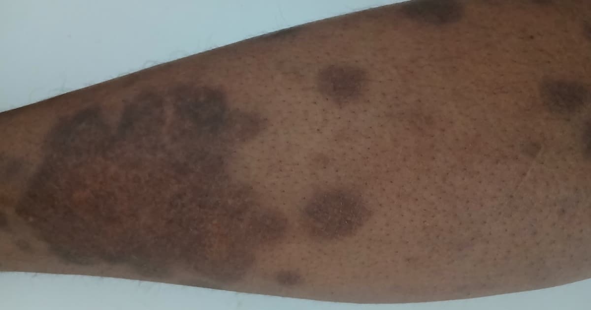



Table of contents
- Why skin AI became both a promise and a risk
- What the Diverse Dermatology Images measured
- Transparency of the datasets
- Fine-tuning and what changes with diverse data
- CLEAR Derm checklist and what to ask the supplier
- What changes in the clinical decision
- Studies at a glance
- How NEXO helps train this reasoning
- Frequently asked questions
- Take-home messages
- References
In a fictional encounter, a skin clinic tests an app that classifies photos of lesions as benign or malignant. The algorithm is often right on the light images in the demonstration set. In the same week, three patients with dark skin tones bring photos of spots and nodules: the app returns low confidence scores or suggests benignity for lesions the dermatologist considers suspicious. The team asks whether the problem is the photo, the device or the model.
This scenario sums up what recent studies have measured: skin image classifiers trained on poorly diverse sets lose performance on dark tones and on less common diseases. This text gathers what the literature shows about that drop, why the datasets fall short on transparency and what the clinician can ask before trusting the result. In each part, the text reports the design and size of the studies and separates what was measured from what is our inference.
Key points
- In the Diverse Dermatology Images set, three leading algorithms had an area under the ROC curve between 0.56 and 0.67, well below the performance reported in their own tests (Daneshjou et al., 2022).
- Sensitivity fell sharply in Fitzpatrick V-VI: ModelDerm from 0.41 to 0.12 and DeepDerm from 0.69 to 0.23 (Daneshjou et al., 2022).
- In 70 AI studies in dermatology, only 24.2 percent of the images were public, 20 percent described race or ethnicity and 10 percent mentioned skin tone (Daneshjou et al., 2021).
- Fine-tuning with diverse images closed the gap between light and dark tones in DeepDerm and in the model trained on HAM10000 (Daneshjou et al., 2022).
- Dermatologists also made more errors on dark tones and on uncommon diseases, which matters because they label many training sets (Daneshjou et al., 2022).
- Why skin AI became both a promise and a risk
- What the Diverse Dermatology Images measured
- Transparency of the datasets
- Fine-tuning and what changes with diverse data
- CLEAR Derm checklist and what to ask the vendor
- What changes in the clinical decision
- Studies at a glance
- How NEXO helps train this reasoning
- Frequently asked questions
- Take-home messages
Why skin AI became both a promise and a risk
Automatic classification of skin lesions gained attention after deep neural networks matched dermatologists on melanoma and carcinoma tasks in large sets (Esteva et al., 2017). The Stanford study trained on 129,450 images and tested on 2,032 biopsy-confirmed images, with performance close to that of 21 board-certified dermatologists (Esteva et al., 2017).
The 2017 leap and the 2018 warning
That milestone sped up apps, teledermatology and the hope of screening in primary care. The following year, Adamson and Smith warned that models trained on poorly representative data can amplify disparities if they are deployed without measuring performance by subgroup (Adamson and Smith, 2018). The warning anticipated what the Diverse Dermatology Images (DDI) measured in numbers years later.
The promise still holds for common lesions in balanced sets, but the clinical question is no longer "is the algorithm right?" and has become "on whom is it right?". This shift in the question connects with the use of artificial intelligence in clinical reasoning and with the risk of trusting the machine too much.
What the Diverse Dermatology Images measured
The DDI was created to test algorithms on clinically confirmed images, with diverse skin tones and common and uncommon diseases (Daneshjou et al., 2022). After excluding photos that failed quality control, 656 images remained, balanced between benign and malignant at the extremes of Fitzpatrick (Daneshjou et al., 2022).
Composition of the set
There were 208 Fitzpatrick I-II images (159 benign and 49 malignant), 241 of III-IV and 207 of V-VI (159 benign and 48 malignant) (Daneshjou et al., 2022). The subsets at the extremes were matched by diagnostic class for the comparison between light and dark tones.
Performance of the three algorithms
In the full DDI, ModelDerm had an area under the ROC curve of 0.65 (95 percent CI 0.61 to 0.70), DeepDerm 0.56 (0.51 to 0.61) and the model trained on HAM10000 0.67 (0.62 to 0.71) (Daneshjou et al., 2022). On their own test sets, these algorithms had reached much higher values, which shows a drop when leaving the original environment.
In the comparison between Fitzpatrick I-II and V-VI, ModelDerm went from 0.64 (0.55 to 0.7) to 0.55 (0.46 to 0.64), DeepDerm from 0.61 (0.50 to 0.71) to 0.50 (0.41 to 0.58) and HAM10000 from 0.72 (0.63 to 0.79) to 0.57 (0.48 to 0.67) (Daneshjou et al., 2022). Sensitivity fell from 0.41 to 0.12 in ModelDerm and from 0.69 to 0.23 in DeepDerm (Daneshjou et al., 2022).
Dermatologists also miss on dark skin
The DDI asked three dermatologists to classify each image as a benign or malignant process and formed a majority vote (Daneshjou et al., 2022). The specialists' labels outperformed the algorithms before fine-tuning, but the dermatologists' sensitivity also fell from 0.72 in Fitzpatrick I-II to 0.59 in V-VI (Daneshjou et al., 2022).
The test with 848 physicians
Groh and colleagues ran a digital experiment with 389 dermatologists and 459 primary care physicians from 39 countries, who assessed 364 images of 46 diseases in a teledermatology simulation (Groh et al., 2024). Diagnostic accuracy was 38 percent among dermatologists and 19 percent among generalists, and both groups were 4 percentage points less accurate on dark skin (Groh et al., 2024). Support from a deep learning algorithm increased accuracy by more than 33 percent, but widened the difference between skin tones among generalists (Groh et al., 2024). The authors conclude that improving overall accuracy does not, by itself, solve the bias (Groh et al., 2024).
Why this matters for training
Many datasets use dermatologists' labels as the reference standard. If human error is greater on dark tones and on uncommon diseases, the algorithm inherits and can amplify that failure (Daneshjou et al., 2022). The discussion intersects with implicit bias in diagnosis and with confidence calibration: accuracy and certainty do not move together when the subgroup changes.
One synthesis of ours is that the dermatologist remains indispensable, but is not a gold standard free of photographic bias; their role is to filter the algorithm, not just to label the dataset.
Transparency of the datasets
Before the DDI, the scoping review in JAMA Dermatology examined 70 deep learning studies in dermatology (Daneshjou et al., 2021). Among 1,065,291 images used to develop or test algorithms, only 257,372 (24.2 percent) were publicly available (Daneshjou et al., 2021).
What almost no one reports
Only 14 studies (20.0 percent) described ethnicity or race in at least one set, and only 7 (10.0 percent) reported any skin tone data (Daneshjou et al., 2021). Without this information, the reader cannot know whether the model was tested in the population where it will be used.
Public datasets and network effect
Public sets were cited more often than private ones, which concentrates training in a few sources and in photographs with a similar demographic profile (Daneshjou et al., 2021). One hypothesis of ours is that the repetition of the same datasets explains part of the drop seen in the DDI when the clinical setting changes.
The scoping review also showed that the median number of images in studies that trained new algorithms was about 10 thousand, with wide variation, which reinforces the inequality of resources between research groups (Daneshjou et al., 2021). Without public data and without demographic description, the clinical community cannot reproduce or audit performance by subgroup before purchase or deployment.
What the public datasets contain
Wen and colleagues systematically reviewed 21 open-access skin cancer image banks, with 106,950 images (Wen et al., 2022). Of the 14 banks that reported the country of origin, 11 (79 percent) came from Europe, North America and Oceania, and 19 banks (91 percent) had only dermoscopic or macroscopic images (Wen et al., 2022). Ethnicity was available for 1,415 images (1.3 percent) and Fitzpatrick phototype for 2,236 (2.1 percent) (Wen et al., 2022). The authors call for quality standards for bank metadata (Wen et al., 2022).
This opacity echoes the warning from the report on diagnostic error: systems that do not measure failure by subgroup repeat inequity, the theme of the text on the Improving Diagnosis report.
Fine-tuning and what changes with diverse data
The DDI authors retrained DeepDerm and the HAM10000 model with diverse images (Daneshjou et al., 2022). After fine-tuning, DeepDerm reached an area under the curve of 0.73 (95 percent CI 0.70 to 0.77) in Fitzpatrick I-II and 0.76 (0.71 to 0.80) in V-VI (Daneshjou et al., 2022).
HAM10000 after tuning
The model based on HAM10000 reached 0.77 (0.73 to 0.80) in I-II and 0.78 (0.75 to 0.81) in V-VI (Daneshjou et al., 2022). The gap between light and dark tones closed, and performance came to match or exceed dermatologists' vote in several data slices.
Reading for the clinician
The result does not mean that any commercial application has already been adjusted. It means that the failure is correctable when there are labeled and diverse images, and that asking for evidence of testing by skin tone is a legitimate question before adoption. The 2025 narrative review reached a similar conclusion: most studies showed worse accuracy in Fitzpatrick IV-VI, with improvement after training on diverse sets (Dowie, 2025).
Skin tone is not the same as race
Fitzpatrick was created to estimate response to ultraviolet light, not to map ancestry. Ulrich and colleagues described an automated method that estimates skin tone and converts it to the Monk and Fitzpatrick scales; in validation with 3D body scans and AI-generated images, agreement was high with Monk and less consistent with Fitzpatrick (Ulrich et al., 2025).
Why the distinction matters in the consultation
Self-reported race, ethnicity and skin tone measured in the photo answer different questions. An algorithm can fail due to pigmentation, lighting, lesion type or a mix of these factors (Daneshjou et al., 2022; Ulrich et al., 2025). Treating "dark skin" as a single category without describing the classification method hides the mechanism of the error.
In practice, the clinician who documents the observed tone and the photo quality helps both the algorithm audit and human reasoning, in the same spirit of avoiding labels that hide the rest of the picture.
CLEAR Derm checklist and what to ask the supplier
The AI working group of the International Skin Imaging Collaboration published CLEAR Derm, a checklist of good practices for development and evaluation of image-based AI in dermatology (Daneshjou et al., 2022). The document covers data curation, model construction and evaluation, with attention to photographic bias and ethical considerations (Daneshjou et al., 2022).
Five practical questions (our synthesis)
Before incorporating a classifier into routine, a list of ours, not validated as an instrument, includes:
- Was performance reported by skin tone or by an equivalent scale?
- Is the test set external to the training set?
- Are there sensitivity and specificity rates by subgroup, not just global area under the curve?
- Are the training images publicly auditable or at least described in demographic composition?
- Is there a plan for human supervision and recording when the algorithm and the clinician disagree?
These questions dialogue with the critical reading of articles and with care in the melanoma team, where the image is just one step along the way.
Lighting, capture and the path to the pixel
The algorithm only sees what the camera delivers. Side lighting, shadow, file compression and a colored background change apparent texture and color. The review of principles and applications of AI in dermatology reinforces that image quality and the representativeness of the database limit any model (Omiye et al., 2023).
What to standardize in the office
Our operational list, based on the limits described by the studies and not tested as a protocol, includes:
- Use diffuse frontal light and avoid harsh flash that blows out the highlight.
- Frame the lesion with a margin of healthy skin around it.
- Keep the same distance and the same angle in follow-up photos.
- Record the device and the date alongside the clinical file.
- Avoid sending blurred photos or photos with a beauty filter to the algorithm.
These precautions also apply to traveler lesions and to differential diagnoses of skin in mpox and in traveler skin, even when the algorithm does not enter the decision.
What changes in the clinical decision
In primary care, this means not delegating the biopsy decision to a score without knowing in which population it was validated. The same care applies when the photo arrives by message: poor quality and a skin tone outside the training database increase uncertainty, and the clinician should state this clearly to the patient.
Faced with an algorithmic score, the clinician still needs the history, the examination and the epidemiological context. One hypothesis of ours is that the greatest risk is not the false positive on light demonstration skin, but the false negative on dark skin when the app suggests "probably benign" with apparent confidence.
How to integrate without automating the closure
Practical suggestions, marked as a synthesis:
- Treat the algorithm result as a clue, not as a report.
- Refuse a decision based only on a score when the skin tone or the disease falls outside the spectrum of the validation study.
- Document disagreement between clinician and machine for future audit.
- Prioritize biopsy or referral when the lesion is clinically suspicious, regardless of the app.
- Avoid announcing to the patient an "artificial intelligence confirmation" without explaining the limits.
This use fits the training of differential diagnosis with clues and reduces diagnostic error in primary care linked to early closure.
When the algorithm and the clinic diverge
If the lesion looks suspicious on examination and the app suggests benignity, management follows the clinic. The DDI showed low sensitivity in Fitzpatrick V-VI precisely in the slices where the false negative hurts most (Daneshjou et al., 2022). In these cases, biopsy, in-person dermoscopy or a second opinion weigh more than the score.
One synthesis of ours is to record in the chart the algorithm result, the observed skin tone and the final decision. This record creates the local audit base that multicenter studies still rarely publish by subgroup.
Limits that the text does not resolve
The DDI numbers come from a curated set at an academic center and do not measure the performance of each commercial app available today. Dowie's narrative review selected few articles after restrictive filters (Dowie, 2025). Ulrich validates a method for classifying tone, not a cancer classifier (Ulrich et al., 2025).
Therefore, generalizing that "all skin AI fails" would be improper inference. What the data support is narrower: algorithms trained on poorly transparent and poorly diverse databases have a measurable risk of worse performance on dark tones, and this risk decreases when training and testing include these images on purpose (Daneshjou et al., 2022; Daneshjou et al., 2021).
What the clinician takes to the next consultation
After reading the DDI and the transparency review, the practical change is not to abandon the technology, but to demand the same rigor required of a new laboratory test. One synthesis of ours, aligned with the findings cited, is to organize the consultation in three steps: (1) quality of the photo and of the observed skin tone; (2) algorithm result as a clue, never as a report; (3) documented decision when there is disagreement.
How to explain the limit to the patient
One formulation of ours, not tested as a script, is to say that the app helps organize hypotheses, but it was trained mostly on images of certain skin tones, and that the decision to biopsy or observe remains clinical. This avoids both blind dismissal of the tool and the automatic trust that automation bias describes.
Minimum audit in the service
For services that already use a classifier, an operational checklist of ours includes reviewing monthly the cases in which the algorithm and the dermatologist disagreed, separating the files by observed skin tone and checking whether the false negatives cluster in Fitzpatrick V-VI, as the DDI predicted (Daneshjou et al., 2022). Without this cycle, the service repeats the opacity that the review of 70 studies documented (Daneshjou et al., 2021).
The same spirit of measurement by subgroup appears in team melanoma care and in preventing premature closure, the theme of differential diagnosis training with clues. Technology only reduces harm if the service knows whom it fails.
Studies at a glance
| Study | Design, sample and unit | Main finding |
|---|---|---|
| Esteva 2017 | Neural network; 129,450 training images, 2,032 test images | Performance close to that of dermatologists on its own dataset |
| Adamson 2018 | Commentary | Warning of disparity if measurement by subgroup is missing |
| Daneshjou 2021 | Scoping review; 70 studies | Little transparency on race and skin tone |
| Daneshjou 2022 (DDI) | External evaluation; 656 images | Drop in AUC and sensitivity in Fitzpatrick V-VI |
| Wen 2022 | Systematic review; 21 public datasets, 106,950 images | Fitzpatrick reported in 2.1 percent of the images (Wen et al., 2022) |
| Groh 2024 | Digital experiment; 848 physicians, 364 images | Dark skin with lower accuracy; AI support widened the gap between generalists (Groh et al., 2024) |
| CLEAR Derm 2022 | Checklist consensus | Items for data, model and fair evaluation |
| Omiye 2023 | Narrative review | Limits of imaging and representativeness |
| Dowie 2025 | Narrative review; 8 final articles | Worse accuracy in dark tones; improvement with diverse data |
| Ulrich 2025 | Tone analysis method | Monk scale with better alignment in some tests |
Sources: Esteva et al., 2017; Adamson and Smith, 2018; Daneshjou et al., 2021; Daneshjou et al., 2022; Daneshjou et al., 2022; Wen et al., 2022; Groh et al., 2024; Omiye et al., 2023; Dowie, 2025; Ulrich et al., 2025.
How NEXO helps train this reasoning
In NEXO, the case of the day is fictional, with a real clinical presentation and up to six progressive clues, and you propose the diagnosis by searching for the ICD-10 code. One clue may say "asymmetric patch on the back, patient with dark skin tone"; another, "skin app suggested benignity with low confidence". The five-level proximity feedback helps separate nevus, melanoma, basal cell carcinoma and dermatitis, without turning the training into a software protocol.
Frequently asked questions
Does every skin app fail on dark skin?
The three current algorithms tested in the DDI dropped in Fitzpatrick V-VI, and later reviews found the same pattern in most studies (Daneshjou et al., 2022; Dowie, 2025).
Does tuning the model with diverse data solve it?
In the DDI, fine-tuning closed the gap between I-II and V-VI in DeepDerm and in the HAM10000 model, but each commercial product must show its own test (Daneshjou et al., 2022).
Are dermatologists immune to photo bias?
The sensitivity of the majority vote fell from 0.72 to 0.59 between light and dark tones in the DDI (Daneshjou et al., 2022).
Are skin tone and race the same variable?
Fitzpatrick describes response to light; race and ethnicity are distinct social categories, and scales such as Monk try to capture the tone observed in the image (Ulrich et al., 2025).
What should be requested before adopting a classifier?
Performance by skin tone subgroup, external testing, sensitivity and specificity, not only the global area under the curve, and a human supervision plan, in the spirit of CLEAR Derm (Daneshjou et al., 2022).
Take-home messages
- Current algorithms lost AUC and sensitivity on dark skin in the DDI (Daneshjou et al., 2022).
- Most AI studies in dermatology report neither race nor skin tone (Daneshjou et al., 2021).
- Fine-tuning with diverse images can close the gap, if there is data and audit (Daneshjou et al., 2022).
- Dermatologists also make more errors on darker tones, and AI support can widen the gap between generalists (Daneshjou et al., 2022; Groh et al., 2024).
- The clinician should treat the score as a clue and demand transparency in the CLEAR Derm style (Daneshjou et al., 2022).
References
- Esteva A, Kuprel B, Novoa RA, et al. Dermatologist-level classification of skin cancer with deep neural networks. Nature. 2017;542(7639):115-118. PMID: 28117445. https://www.ncbi.nlm.nih.gov/pmc/articles/PMC8382232/
- Adamson AS, Smith A. Machine Learning and Health Care Disparities in Dermatology. JAMA Dermatol. 2018;154(11):1247-1248. PMID: 30073260. https://pubmed.ncbi.nlm.nih.gov/30073260/
- Daneshjou R, Smith MP, Sun MD, et al. Lack of Transparency and Potential Bias in Artificial Intelligence Data Sets and Algorithms: A Scoping Review. JAMA Dermatol. 2021;157(11):1362-1369. PMID: 34550305. https://www.ncbi.nlm.nih.gov/pmc/articles/PMC9379852/
- Daneshjou R, Vodrahalli K, Novoa RA, et al. Disparities in dermatology AI performance on a diverse, curated clinical image set. Sci Adv. 2022;8(32):eabq6147. PMID: 35960806. https://www.ncbi.nlm.nih.gov/pmc/articles/PMC9374341/
- Daneshjou R, Barata C, Betz-Stablein B, et al. Checklist for Evaluation of Image-Based Artificial Intelligence Reports in Dermatology: CLEAR Derm Consensus Guidelines from the International Skin Imaging Collaboration Artificial Intelligence Working Group. JAMA Dermatol. 2022;158(1):58-66. PMID: 34851366. https://www.ncbi.nlm.nih.gov/pmc/articles/PMC9845064/
- Omiye JA, Gui H, Daneshjou R, et al. Principles, applications, and future of artificial intelligence in dermatology. Front Med (Lausanne). 2023;10:1278232. PMID: 37901399. https://www.ncbi.nlm.nih.gov/pmc/articles/PMC10602645/
- Dowie T. Exploring the Diagnostic Capability of Artificial Intelligence in Dermatology for Darker Skin Tones: A Narrative Review. Cureus. 2025;17(10):e94909. PMID: 41262830. https://www.ncbi.nlm.nih.gov/pmc/articles/PMC12624499/
- Ulrich P, Zink A, Biedermann T, Sitaru S. Beyond Fitzpatrick: automated artificial intelligence-based skin tone analysis in dermatological patients. NPJ Digit Med. 2025;8:378. PMID: 40537526. https://www.ncbi.nlm.nih.gov/pmc/articles/PMC12179258/
- Wen D, Khan SM, Ji Xu A, et al. Characteristics of publicly available skin cancer image datasets: a systematic review. Lancet Digit Health. 2022;4(1):e64-e74. PMID: 34772649. https://pubmed.ncbi.nlm.nih.gov/34772649/
- Groh M, Badri O, Daneshjou R, et al. Deep learning-aided decision support for diagnosis of skin disease across skin tones. Nat Med. 2024;30(2):573-583. PMID: 38317019. https://www.ncbi.nlm.nih.gov/pmc/articles/PMC10878981/