Retained bullet: when to remove it and lead risk
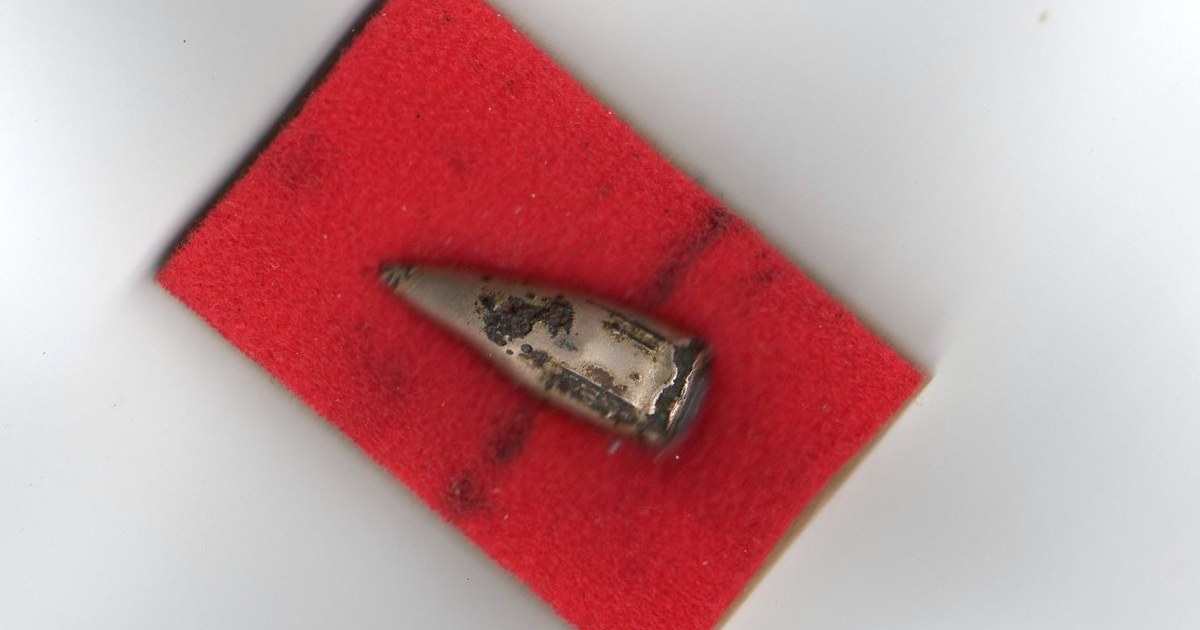
Table of contents
The following scene is fictional and was written for teaching. A 50-year-old man comes to the outpatient clinic with cramping abdominal pain, constipation, fatigue and weight loss for two months. He has already had an endoscopy and an ultrasound, with no findings. On examination, he is pale. When asked about his history, he recalls that he was shot in the hip almost twenty years ago and that the bullet "stayed in there". The blood count shows mild anemia. The hip X-ray shows metal fragments inside the joint. Can a bullet forgotten two decades ago explain the abdominal pain?
This text summarizes what happens to bullets that stay in the body after a firearm injury: when to leave them, when to remove them, the risk of lead poisoning and migration through blood vessels. The content is educational, aimed at students and health professionals, and does not replace local protocols or the assessment of surgeons, toxicologists and poison information centers.
Key points
- Most retained bullets are well tolerated and do not need to be removed.
- The risk of lead poisoning is low, except when the bullet is in contact with synovial fluid, an intervertebral disc or cerebrospinal fluid.
- The symptoms of poisoning are nonspecific and may appear years or decades after the injury.
- Blood lead level measurement is the central test for investigating the suspicion.
- Treatment combines removal of the source, when possible, and chelation led by a specialist.
- Bullets can migrate through blood vessels to the heart, the lungs or a limb, and this requires individual assessment.
Why many bullets stay
After a firearm injury, the bullet, or its fragments, often stays in the body. Searching for every fragment would require dissecting healthy tissue, would increase bleeding and the risk of infection and, in many cases, would cause more harm than the bullet itself. Therefore, the rule in the acute phase is to treat the injuries and leave the metal that does not cause trouble.
An American review on ballistics, pathology and retained bullets notes that, contrary to what is often thought, the risk of lead poisoning is uncommon unless the bullet is in contact with synovial fluid or an intervertebral disc (Baum et al., 2022). The same review points out that existing guidelines are largely old and that, according to a 2022 survey, only 14.5 in every 100 surgeons reported having institutional policies for bullet removal.
The lack of clear rules means the decision depends on each team's judgment. This makes it even more important that the patient knows they have a retained bullet, where it is and which symptoms should lead them to seek care. The information must appear in the discharge summary and be passed on to the doctor who will provide follow-up.


When to remove
The decision to remove a bullet considers its location, the symptoms, the risk of surgery and the risk of leaving the metal. There is no universal list, but the situations below appear frequently in the literature reviewed in this text:

- Bullet inside a joint, due to the risk of lead poisoning and arthritis.
- Bullet in contact with cerebrospinal fluid or inside the intervertebral disc.
- Bullet inside a blood vessel or a heart chamber, due to the risk of migration and embolism.
- Bullet that compresses a nerve and causes pain or deficit.
- Bullet associated with infection, abscess or fistula.
- Superficial bullet, palpable and causing pain or discomfort.
- Clinical or laboratory signs of lead poisoning.
Even in these cases, the timing can vary. In an unstable patient, survival is the priority, and the projectile can be removed later. In a stable patient with an intra-articular projectile, many surgeons prefer early removal, before the lead dissolves in the synovial fluid, usually by arthroscopy or a small arthrotomy.
The review by Baum and colleagues cites a case of projectile removal from the ankle 49 years after the injury, due to arthropathy and systemic lead poisoning (Baum and colleagues, 2022). The example shows that the risk does not disappear with time.
Lead inside the body
Most projectiles have a lead core, with or without a coating of copper or another alloy. When the projectile becomes surrounded by fibrous tissue, in muscle or fat, the fibrosis capsule isolates the metal and little lead is absorbed. In fluids such as synovial and cerebrospinal fluid, this capsule does not form well, and the lead slowly dissolves. Baum and colleagues explain that lead is highly soluble in synovial fluid, and that projectiles retained in joint spaces can cause local and systemic effects (Baum and colleagues, 2022).
A case-control study in Rio de Janeiro compared 45 victims of gunshot wounds with metal fragments retained for more than 6 months with 45 controls matched by sex, age and race. The mean blood lead level was 9.01 micrograms per deciliter in the cases and 2.17 in the controls. The cases more often reported irritability, bad mood, headache, memory loss, daytime drowsiness, muscle pain, weakness, abdominal pain, joint pain, tremor and tingling (de Araújo and colleagues, 2015).
The authors concluded that retained projectiles are not harmless, with effects on blood lead levels and on symptoms even with few fragments, location outside the joint or low lead levels. They recall the World Health Organization position that no level of lead exposure is considered safe (de Araújo and colleagues, 2015). The finding does not mean that every projectile should be removed, but it reinforces the importance of follow-up.
Factors often associated with greater lead absorption from a retained projectile:
- Location in a joint, intervertebral disc or space with cerebrospinal fluid.
- Fragmentation, which increases the metal's contact surface.
- Associated fracture, with contact of the metal with bone undergoing remodeling.
- Long dwell time.
- Situations that increase bone remodeling, such as pregnancy, immobilization and hyperthyroidism.
- Infection or inflammation around the projectile.
Symptoms of lead poisoning
Lead poisoning is called the great imitator because of its nonspecific symptoms. Baum and colleagues describe that symptoms such as headache, fatigue, nausea, abdominal discomfort, anemia, kidney failure, encephalopathy and neuropathy usually appear only with serum levels above 24 micrograms per deciliter (Baum and colleagues, 2022).
Abdominal pain is one of the most misleading symptoms. One report describes a 48-year-old man with abdominal pain, weight loss and constipation, with a history of a gunshot wound to the left hip about 20 years earlier. The radiograph showed numerous intra-articular fragments and calcified hemarthrosis around the femoral head. The blood lead level was raised, and chelation led to improvement of symptoms (Abraham and colleagues, 2012). The authors note that the unpredictable latency period and the nonspecific symptoms make diagnosis difficult, and that poisoning can be fatal if not recognized.
An older report describes a 23-year-old man with abdominal cramps, weakness, vomiting and diarrhea with dark stools, recurring for two years. The radiograph showed a projectile lodged in the left hip, with the natural contrast of the lead outlining the entire synovial capsule (de Madureira and colleagues, 2000). After chelation, the symptoms returned, and the solution was complete replacement of the hip joint with a prosthesis.
| System | Possible manifestations |
|---|---|
| Gastrointestinal | Cramping abdominal pain, constipation, nausea, vomiting, weight loss |
| Hematologic | Anemia, basophilic stippling in red blood cells |
| Central nervous | Headache, irritability, memory impairment, encephalopathy |
| Peripheral nervous | Tingling, weakness, motor neuropathy |
| Renal | Kidney injury, which may be progressive |
| Musculoskeletal | Joint pain, arthropathy in the joint where the projectile is located |
Returning to the scene from the beginning, the combination of cramping abdominal pain, constipation, anemia and metal fragments inside the hip looks very much like the reports described above. The hypothesis of lead poisoning does not rule out other causes, and the abdominal investigation continues, but the blood lead level becomes a priority test.
Diagnosis and follow-up
The blood lead level is the central test. In patients with an intra-articular projectile, in contact with cerebrospinal fluid or with compatible symptoms, it can be repeated over time to detect a rise. A report on poisoning after a comminuted knee fracture with retained fragments highlights that stopping the exposure is the main focus, and that monitoring lead levels and chelation are part of management when complete removal of the fragments is not feasible (Costantini et al., 2023).
Other tests help assess the impact of lead: complete blood count with smear, kidney function, erythrocyte protoporphyrin and, in some services, urinary aminolevulinic acid. Radiography shows the position and fragmentation of the projectile, and CT helps define its relationship with joints and with the spinal canal. Baum et al. note that MRI is contraindicated because common projectiles are metallic (Baum et al., 2022), and the decision to perform the exam depends on a case-by-case assessment by the radiologist.
Long-term follow-up has no standardized schedule. A reasonable strategy, discussed in the reviewed reports, is to measure lead when symptoms appear, after surgeries that involve the region of the projectile and in higher-risk situations, such as new fractures in the same bone. Changes in the radiographic pattern, such as the outline of the joint capsule drawn by the metal, also draw attention to lead dissolution.
Diagnosis depends heavily on remembering the projectile. In patients with unexplained abdominal pain, anemia, neuropathy or cognitive changes, asking about old gunshot wounds, even decades earlier, can shorten a long investigation.
Treatment of poisoning
Treatment has two pillars: removing the source and reducing the body's lead burden. When the projectile is accessible, removal is the most important step, because chelation without removal tends to be followed by relapse. The report by Costantini et al. highlights the lack of guidelines for cases in which complete removal is not feasible, the importance of a multidisciplinary decision and respect for patient autonomy (Costantini et al., 2023).
Chelation uses drugs that bind to lead and help its elimination in urine. In the reports reviewed here, intravenous calcium disodium edetate (de Madureira et al., 2000) and oral succimer (Abraham et al., 2012) were used. The choice of drug, dose and duration should be defined with a toxicologist or a poison information center, according to the lead level, symptoms and kidney function.
Another case shows how treatment can last for years. A 42-year-old man had a projectile retained next to the spinal cord after a robbery. Years later, he developed severe low back pain and underwent surgery twice; after the second surgery, severe abdominal pain appeared. For five years, he received cycles of chelation with calcium edetate, with a good response during symptomatic periods, until the markers normalized (Madureira et al., 2009). The authors linked the poisoning to contact between the projectile and the cerebrospinal fluid.


Projectiles that migrate
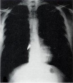
Projectile embolism happens when the projectile, or a fragment, enters a vessel without leaving it and is carried by the blood. In the venous circulation, it travels to the right heart and the pulmonary arteries; in the arterial circulation, to the brain, the viscera or the limbs. One report describes a 19-year-old with multiple wounds to the trunk whose projectile ended up in the left popliteal artery, requiring embolectomy. The investigation led to the diagnosis of a false aneurysm of the abdominal aorta that had not been clear before (Huang et al., 2016). The authors note that peripheral embolism is often misdiagnosed for lack of early symptoms.
Another report describes a 29-year-old man struck in the left arm, with the projectile entering the chest without an exit. The initial radiograph showed the projectile in the left thigh, and the CT scan revealed a false aneurysm of the descending thoracic aorta and the projectile inside the superficial femoral artery (Pereira et al., 2018). The patient had diminished pulses in the left foot and numbness in the toes, a clue that the projectile was in the wrong place. The aortic false aneurysm was treated with a stent graft, and the projectile was removed through an incision in the left groin, with embolectomy (Pereira et al., 2018). The authors stress that the diagnosis depends on a high degree of suspicion.
In the venous circulation, one report describes projectile embolism to the pulmonary artery with pulmonary infarction after a wound to the left eye (Duke et al., 2014). Another report, on venous embolism to the right ventricle, recommends surgical removal of projectiles in the right heart when the risk of sternotomy and cardiotomy is low, and considers endovascular removal as an alternative. The review lists three options, observation, endovascular removal and surgical removal, and acknowledges that there is controversy, because there are several reports of success with observation alone. The authors recommend transesophageal echocardiography before any attempt to remove a foreign body in the heart (Henderson et al., 2021).
Signs that suggest projectile embolism:
- Entry wound without an exit and without a projectile along the expected path.
- Projectile in a location that is anatomically impossible from the wound.
- Projectile that changes position between exams.
- Limb or visceral ischemia far from the wound.
- New neurological deficit.
- Chest pain, shortness of breath or arrhythmia without direct chest injury.
Spine, lung and other locations
Projectiles in the spinal canal are a special problem. A report of a projectile retained in the cervical spinal canal, with complete spinal cord injury at the C4 level, describes the doubts about surgery in these cases: removal rarely improves neurological function, and the decision depends on several factors. The review discusses that early removal may prevent lead toxicity from contact with the cerebrospinal fluid (Benton et al., 2020). The patient in that report was treated without surgery, remained ventilator-dependent and died of cardiac arrest attributed to pulmonary embolism.
In the lung, most projectiles are left in place. An Iraqi report describes a 46-year-old man with vague chest pain at the site of a wound from 26 years earlier, with a 22-millimeter foreign body in the left lower lobe. The projectile was removed by uniportal video-assisted thoracoscopy, and the defect was sutured (Kakamad et al., 2021). Minimally invasive techniques have made removal simpler when there is an indication.
The review by Huang et al. recalls that embolism may follow the arterial or venous system, with common sites that include the left ventricle, the pulmonary vein, the thoracic and abdominal aorta and the peripheral arteries (Huang et al., 2016). Therefore, the search for a projectile that does not appear along the path should include the chest, abdomen and limbs, and not only the region of the wound. A radiograph that does not find the projectile where it should be is an important finding, not a detail.
In the limbs, projectiles in soft tissues are usually left, unless they lodge in a joint, cause pain or compress a nerve. Projectiles in bones may be removed during fracture fixation, when accessible. In all locations, follow-up includes advising the patient about warning symptoms and recording the presence of the metal in the medical record.
What blood lead surveillance showed
A report from the United States Centers for Disease Control and Prevention analyzed adults with blood lead at or above 10 micrograms per deciliter reported to the national surveillance program between 2003 and 2012. There were 145,811 people aged 16 and older, in 41 states, and 457 of them had retained bullet fragments (Weiss et al., 2017). The highest value linked to a retained fragment was 306 micrograms per deciliter (Weiss et al., 2017).
The numbers show the size and the profile of the problem. Cases linked to bullets accounted for 0.3 percent of all raised levels, but 4.9 percent of levels above 80 micrograms per deciliter, meaning they weigh more among severe poisonings (Weiss et al., 2017). They occurred mostly in men aged 16 to 24, outside the workplace (Weiss et al., 2017).
The report also explains why this diagnosis slips by. Blood lead testing in adults is usually ordered to monitor occupational exposure, and routine testing in people with a retained fragment is uncommon. Symptoms are nonspecific, such as fatigue, abdominal pain and memory loss, and can appear years after the injury (Weiss et al., 2017). Until 2004, fewer than one hundred reports of lead poisoning from retained bullets had been published in the medical literature, which suggests that surveillance finds far more cases than isolated reports reveal (Weiss et al., 2017).
For clinical reasoning, the message is direct. When faced with anemia, abdominal pain without a clear cause, neuropathy or memory changes, asking about old gunshot wounds can shorten the investigation, and blood lead testing confirms or rules out the hypothesis.
How NEXO helps train this reasoning
Training with fictional clinical cases helps you recognize patterns before you meet them on shift, as long as each clue is read calmly and compared with the hypotheses that are still open. Lead poisoning from a retained bullet is an example of a diagnosis that depends on a clue from the past: the abdominal pain and anemia only make sense when the old wound comes up. The habit of reviewing the case before concluding is covered in the text on diagnostic pause, and hidden injuries in the text on blast trauma.
The texts on differential diagnosis with clues, diagnostic uncertainty and clinical decision-making also help. To understand points and rewards, see the NEXO economy page. There are also tracks for those who research (Researcher), for those who review cases (Curator), for those who produce content (Creators) and for those who want to follow results (Closer).
Frequently asked questions
Is having a bullet in the body dangerous?
Most of the time, no. Bullets surrounded by fibrosis in muscle or fat rarely cause problems. The risk increases when the bullet is in a joint, an intervertebral disc, the spinal canal or inside a vessel.
How long after can poisoning appear?
Months, years or decades. The reports reviewed in this text describe cases with 20 years and even 49 years between the injury and the diagnosis.
Should people with a retained bullet have their blood lead tested?
Testing is indicated when there are compatible symptoms or when the bullet is in a risky location, such as a joint or in contact with cerebrospinal fluid. For the others, management follows the local protocol.
Do small fragments also release lead?
Yes. Fragments increase the metal's contact surface with tissues, and the case-control study cited in this text found higher lead levels and more symptoms even in patients with few fragments and outside the joints. This does not require removal, but it justifies attention to symptoms.
Does chelation solve it without removing the bullet?
It lowers blood lead, but if the source remains, levels tend to rise again. When possible, removing the bullet is the main step.
Can I have an MRI with a bullet in the body?
It depends on the bullet's material and location. Many services contraindicate the exam, and the decision must be made by the radiologist based on the imaging and the history.
Can a bullet move years later?
It can, although it is rare. Projectiles inside vessels, cardiac chambers or fluid-filled spaces such as the spinal canal are more likely to change position. Comparison radiographs help confirm migration when new symptoms appear.
Does pregnancy change anything?
Women with retained projectiles in a risky location should tell their prenatal care team. Lead crosses the placenta, and decisions about testing and follow-up should be made with the obstetrician and, if needed, a toxicologist.
Take-home messages
- Record the presence and position of retained projectiles in the medical chart and in the discharge summary.
- Assess removal of projectiles in a joint, spinal canal, vessel or heart.
- Ask about old wounds in patients with abdominal pain, anemia or unexplained neuropathy.
- Measure blood lead when there is clinical suspicion or a risky location.
- Conduct chelation with a toxicologist or a poison information center.
- Look for embolism when the projectile is not where it should be.
- Instruct the patient about warning symptoms.
If you like reasoning from clues, today's case is waiting on the NEXO home page.
References
- Abraham A, Singh J, Mustacchia P, et al. Pain from a Bullet Lingers on: An Uncommon Case of Lead Toxicity. Case Rep Gastroenterol. 2012. PMID 22679412.
- Baum GR, Baum JT, Hayward D, et al. Gunshot Wounds: Ballistics, Pathology, and Treatment Recommendations, with a Focus on Retained Bullets. Orthop Res Rev. 2022. PMID 36090309.
- Benton JA, Rahme R, Krystal J, et al. Retained bullet in the cervical spinal canal and the associated surgical management conundrum: case report and review of the literature. Spinal Cord Ser Cases. 2020. PMID 32826864.
- Costantini I, Carollo M, Losso L, et al. "Heavy Metal": Management of lead toxicity following a gunshot injury with retained lead fragments, a case report. Toxicol Rep. 2023. PMID 38021473.
- de Araújo GC, Mourão NT, Pinheiro IN, et al. Lead Toxicity Risks in Gunshot Victims. PLoS One. 2015. PMID 26509498.
- de Madureira PR, De Capitani EM, Vieira RJ. Lead poisoning after gunshot wound. Sao Paulo Med J. 2000. PMID 10810333.
- Duke E, Peterson AA, Erly WK. Migrating bullet: A case of a bullet embolism to the pulmonary artery with secondary pulmonary infarction after gunshot wound to the left globe. J Emerg Trauma Shock. 2014. PMID 24550629.
- Henderson L, Wachsman A, Chikwe J, et al. Venous bullet embolism to the right ventricle: Case report and review of management. Clin Case Rep. 2021. PMID 33598272.
- Huang J, Pandey V, Shah R, et al. Popliteal artery embolism of bullet after abdominal gunshot wound. Radiol Case Rep. 2016. PMID 27920844.
- Kakamad FH, Ali RK, Mohammed SH, et al. Single port video-assisted thoracoscopic surgery for removal of a retained bullet in the lung parenchyma; A case report with literature review. Ann Med Surg (Lond). 2021. PMID 34840741.
- Madureira PR, De Capitani EM, Vieira RJ, et al. Lead poisoning due to gunshot bullet in contact with cerebrospinal fluid: case report. Sao Paulo Med J. 2009. PMID 19466296.
- Pereira RM, Souza JEDS, de Araújo AO, et al. Arterial bullet embolism after thoracic gunshot wound. J Vasc Bras. 2018. PMID 30643515.
- Weiss D, Tomasallo CD, Meiman JG, et al. Elevated Blood Lead Levels Associated with Retained Bullet Fragments, United States, 2003-2012. MMWR Morb Mortal Wkly Rep. 2017. PMID 28182606.