Melioidosis: sepsis, pneumonia and abscesses
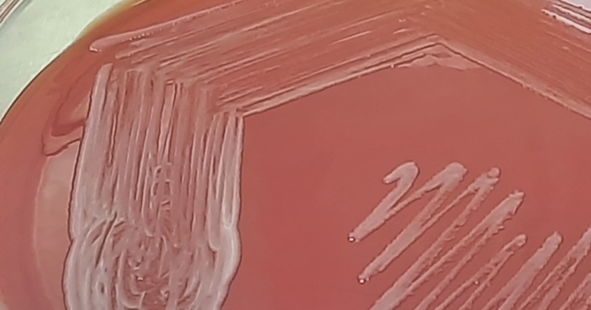
Table of contents
- Why melioidosis is a reasoning setting
- The bacterium, the soil and the rain
- Who is at higher risk
- How the disease presents
- The laboratory: culture, contaminant and warning
- Why the usual antibiotic may fail
- Treatment in two phases
- Relapse, reinfection and adherence
- Endemic regions beyond Southeast Asia
- Reasoning errors in melioidosis
- How NEXO helps train this reasoning
- Frequently asked questions
- Final checklist
- References
In a regional hospital, a farmer in his fifties arrives with high fever for a week, cough and fatigue. He has had diabetes for years, with irregular control, and worked in a flooded rice field right after a season of heavy rains. The X-ray shows pneumonia, he has low blood pressure and the team starts the usual antibiotic for severe pneumonia. Two days later, the blood culture grows a bacterium that the laboratory at first thinks is a contaminant. The scene is fictional and was written for teaching. This text uses that picture as a setting for clinical reasoning: who is at risk of melioidosis, how the disease presents, why the laboratory needs to be warned and why treatment has two phases.
NEXO is a daily medical diagnosis game: one clinical case a day, up to six progressive clues, and ICD-10 proximity feedback across five levels.
The text is not a treatment protocol. Choice of antibiotics, doses, adjustment for kidney function and notification follow national guidelines and the guidance of each service. The interest here is the reasoning: how a question about work and rain changes the hypothesis of a pneumonia that seemed ordinary. If you play the case of the day in NEXO, you will recognize the move: the context clue can be worth as much as the examination clue.
Key points
- Melioidosis is caused by the bacterium Burkholderia pseudomallei, which lives in the soil and water of tropical regions.
- Diabetes is the main risk factor, and most people who fall ill have some risk factor.
- The most common presentation is pneumonia, often with bacteremia and, sometimes, septic shock.
- Abscesses in the spleen, liver, prostate and other organs are important clues.
- Culture is the basis of diagnosis, and the laboratory must be warned of the suspicion.
- Treatment has an intensive intravenous phase and an oral eradication phase, which reduces relapses.
Why melioidosis is a reasoning setting
Melioidosis is usually remembered as a disease of Southeast Asia and northern Australia. Limmathurotsakul and colleagues showed, in a global model, that it is underreported and probably endemic in several countries that have never reported cases, and that the use of ineffective antibiotics contributes to the high lethality (Limmathurotsakul and colleagues, 2016).
Wiersinga and colleagues, in a broad review, describe the disease as "the great imitator", because the signs are nonspecific and can resemble tuberculosis, other pneumonias or sepsis caused by other bacteria (Wiersinga and colleagues, 2018). Anyone who wants a shorter clinical review can read the text by Wiersinga, Currie and Peacock in NEJM.
This is the core of the setting. The disease exists in places where few people think of it, and effective treatment depends on thinking of it early. Severe pneumonia in a patient with diabetes who works with soil and water calls for an extra question. The post on base rate in diagnosis shows how the local frequency of a disease changes the weight of each clue.
The bacterium, the soil and the rain
Burkholderia pseudomallei lives in the soil and surface water of tropical regions. Wiersinga and colleagues describe three routes of entry: through the skin, through small cuts and wounds in contact with soil or water; by inhalation, especially during heavy rains and storms; and by ingestion of contaminated water (Wiersinga and colleagues, 2018).
Work in rice fields and other activities with direct contact with the soil increase exposure. Storms and heavy rains do too, which explains why cases cluster in the rainy season. In the Darwin study, which followed patients for three decades in northern Australia, cases were clearly linked to the rainy season (Currie and colleagues, 2021).
The time between exposure and disease
The incubation period described by Wiersinga and colleagues averages about nine days, ranging from one to twenty-one days. But the bacterium can also stay latent and the disease can appear years after exposure, sometimes when the person already lives far from the endemic region. This possibility makes the history of travel and previous residence as important as recent exposure.


Who is at higher risk
Diabetes is the main risk factor. Wiersinga and colleagues report that about four in five patients with melioidosis have some risk factor (Wiersinga and colleagues, 2018). In the Darwin study, diabetes and harmful alcohol use were among the most common factors (Currie and colleagues, 2021).
Other factors cited in the literature include chronic kidney disease, chronic lung disease and the use of medicines that reduce immunity. For reasoning, the lesson is simple: the more risk factors and the more exposure, the more the hypothesis deserves to be on the list.
The question missing from the history
In the opening scene, diabetes was already in the chart. What changes the hypothesis is the question about work and rain. A history that records only "farmer" loses the information that contact with soil was intense and recent. The post about signs, symptoms and history discusses how to turn social information into a clinical clue.
Useful questions include: what is your job, whether you had contact with mud or standing water, whether there was a storm or flood in the previous weeks, where you were born and where you have lived. Each answer can raise or lower the probability of the hypothesis.
How the disease presents
Melioidosis can range from a localized skin infection to severe sepsis. Wiersinga and colleagues report that about half of patients have pneumonia, that bacteremia is common and that about one in five develops septic shock (Wiersinga and colleagues, 2018). In the Darwin study, pneumonia was also the most frequent presentation (Currie and colleagues, 2021).
Abscesses as a clue
Abscesses are a hallmark of the disease. They can appear in the spleen, liver, prostate, kidneys and, in children from some regions, in the parotid gland. The review by Wiersinga and colleagues recommends considering melioidosis in the presence of any fever in an endemic area, especially with abscesses or pneumonia.
This changes how we read findings that, in isolation, would seem common. A prostate abscess in a man with diabetes who works in the fields, or several small abscesses in the spleen in a person with prolonged fever, should raise the hypothesis.
The neurological form
The review also describes a neurological form, with inflammation of the brain and spinal cord, which can arise after infection through the nose or throat, or through the skin of the limbs. It is rare but severe, and it changes the choice of antibiotic in the intensive phase: meropenem is the preferred drug when there is central nervous system infection (Wiersinga and colleagues, 2018). Limb weakness or cranial nerve changes in a febrile patient from an endemic area deserve this hypothesis.
The chronic form
The chronic form is defined as an infection with symptoms for more than two months. It can resemble tuberculosis, with cough, weight loss and lung lesions, and is often confused with it. The post about differential diagnosis with clues helps keep both hypotheses on the table until the culture decides.
The laboratory: culture, contaminant and warning
Culture is the basis of diagnosis. Wiersinga and colleagues recall that Burkholderia pseudomallei is never part of the normal human flora, and that isolation in any clinical sample should be treated as a diagnosis of melioidosis (Wiersinga and colleagues, 2018).
Even so, the bacterium can be dismissed as a contaminant or wrongly identified as another bacterium, such as Pseudomonas or Bacillus, when the laboratory team is not familiar with it. For this reason, the review recommends that the physician warn the laboratory when melioidosis is suspected.
Why warn the laboratory
There are two reasons. The first is diagnostic: with the warning, the laboratory can use selective media, observe the plates daily for up to four days and use supporting methods, such as latex agglutination and identification by mass spectrometry. The second is safety: the bacterium is classified as a risk group three pathogen, and handling it requires specific precautions.


Which samples to collect
Wiersinga and colleagues point out that blood cultures are the most important sample, because bacteremia is common. The review also recommends, in every suspected case, culture of a throat or rectal swab and of the sediment of centrifuged urine, because these may be the only positive samples in some patients. Pus from abscesses and other samples from the site of infection complete the investigation (Wiersinga and colleagues, 2018).
In practice, this means ordering more than usual in a pneumonia. A urine sample, which would seem irrelevant in a respiratory picture, may be the one that confirms the diagnosis.
The safety pin appearance
Gram staining may show bacilli with stronger staining at the ends, an appearance compared to a safety pin. The review by Wiersinga and colleagues notes that this finding is not reliable for diagnosis: it may be absent and it may appear in other bacteria. It is a clue, and culture remains the standard.


This part of the scenario illustrates what the post about pretest probability discusses: the value of a result depends on who is looking and on what was expected to be found. A "strange" colony in a blood culture carries another weight when the doctor has already told the laboratory what to look for.
Why the usual antibiotic may fail
Burkholderia pseudomallei is naturally resistant to several antibiotics used routinely for pneumonia and sepsis. Limmathurotsakul and colleagues point to the use of ineffective antibiotics as one of the reasons for the high estimated lethality (Limmathurotsakul and colleagues, 2016).
In the opening scene, the usual antibiotic for severe pneumonia may not cover the bacterium. Therefore, in an endemic area and with risk factors, the suspicion should influence the empirical choice before the culture result, according to the local protocol. The post about clinical decision-making shows how to weigh the risk of erring on each side when the decision cannot wait.
The Darwin study and the value of thinking early
The Darwin study followed patients for thirty years and described the fall in mortality over time, attributed to faster diagnosis and access to intensive care (Currie and colleagues, 2021). The lesson for reasoning is that knowing the disease locally, with attentive teams and laboratories, changes the outcome.
For the severe patient, this means treating sepsis with the same care as any severe sepsis: recognize shock early, ensure organ support and transfer to intensive care when necessary, while the right antibiotic takes effect. The appropriate antibiotic and intensive support go together, and neither replaces the other. In small services, arranging in advance with the regional referral how to transfer a patient with suspected melioidosis saves time when the case appears.
Treatment in two phases
Treatment of melioidosis is divided into two phases. The table summarizes the structure described by Wiersinga and colleagues and by later clinical trials. Doses and adjustments follow each country's protocol.
| Phase | Goal | Reference drugs | Described duration |
|---|---|---|---|
| Intensive | Control the acute infection | Intravenous ceftazidime, or meropenem in intensive care or neurological disease | At least ten to fourteen days, more in deep abscesses and complicated infections |
| Eradication | Reduce the risk of relapse | Oral sulfamethoxazole with trimethoprim | At least three months; a trial favored twelve weeks |
The intensive phase
Wiersinga and colleagues describe intravenous ceftazidime as the backbone of the intensive phase, with meropenem reserved for patients in intensive care or with neurological disease, for at least ten to fourteen days and for longer in deep abscesses, complicated pneumonia, neurological disease and bone and joint infections (Wiersinga and colleagues, 2018). The review notes that the fever takes time to subside, with a median of up to nine days, and that this slowness is expected.
This detail matters for the reasoning. A fever that persists for a week on the right antibiotic may be the expected course of the disease. Before switching the regimen, it is worth looking for abscesses that need drainage.
Drainage
The review describes that large single abscesses in the liver and muscles and prostate abscesses usually need drainage, while multiple small abscesses in the spleen, liver and kidneys usually respond to medical treatment. Septic arthritis generally calls for drainage and lavage.
The eradication phase
After the intensive phase comes an oral phase with trimethoprim-sulfamethoxazole. Anunnatsiri and colleagues compared twelve and twenty weeks of this phase in a clinical trial and recommended twelve weeks (Anunnatsiri and colleagues, 2021). Chetchotisakd and colleagues showed that trimethoprim-sulfamethoxazole alone was not inferior to the combination with doxycycline and had fewer adverse effects (Chetchotisakd and colleagues, 2014). A network meta-analysis by Anothaisintawee and colleagues gathered the data on treatments for both phases (Anothaisintawee and colleagues, 2023).
Wiersinga and colleagues note the adverse effects to watch for in this phase, such as skin rash, increased potassium and increased creatinine.
Relapse, reinfection and adherence
The eradication phase exists because the disease can come back. Wiersinga and colleagues explain that recurrence can be a relapse of the same infection or a reinfection from new exposure (Wiersinga and colleagues, 2018). Completing the oral phase reduces the risk of relapse.
Talking about the long phase
Three months of tablets after leaving the hospital is a hard task, especially for those who feel well and go back to work. The conversation at discharge should explain why the oral phase matters, which adverse effects to watch for and when to return for tests. The post on prognostic reasoning shows how talking about what comes after the diagnosis helps the patient plan ahead.
Prevention for those who remain exposed
Wiersinga and colleagues cite Thailand's guidance for residents, farmers and visitors: wear boots and gloves when contact with soil or water is necessary, drink only bottled or boiled water, avoid exposure to heavy rain and dust clouds, stop smoking and avoid applying herbs or organic substances to wounds. The review notes that the effectiveness of these measures still needs more studies, but they are simple and reasonable.
Endemic regions beyond Southeast Asia
The global model by Limmathurotsakul and colleagues estimated that the bacterium is present in the soil of many tropical and subtropical countries where the disease is almost never reported (Limmathurotsakul and colleagues, 2016). The known map of melioidosis therefore shows more where there are laboratories and surveillance than where the bacterium actually lives.
An example in the Americas
South America illustrates this point. Rolim and colleagues reviewed the confirmed cases in the region and reported human cases in Ecuador, Venezuela, Colombia, Brazil and Peru, with the bacterium isolated from the environment in some of these countries. The authors call for greater attention from health authorities and coordinated strategies among countries (Rolim and colleagues, 2018).
A series of seven patients treated at a public hospital in northeastern Brazil, described by Velôso and colleagues, presented fever, cough, pneumonia and abdominal pain as the most common presentations, and the authors concluded that the disease is emerging in the country and that timely diagnosis and treatment can save lives (Velôso and colleagues, 2023). It is a picture that repeats in other areas where the disease is beginning to be recognized.
What local surveillance allows
Where there is organized surveillance, there are teams that know the disease, laboratories that can recognize the bacterium and a clear notification route. Where it is lacking, the same suspicion may depend on the initiative of a single professional who asked the right question. For anyone working in any tropical zone, the hypothesis deserves a place on the list of severe pneumonias and sepsis in people with diabetes, especially after periods of rain.
Outside the endemic zone
Because the bacterium can remain latent, the disease can also appear in people who lived or traveled to endemic regions and now live far from them. The question about where the person has lived is what links the current condition to the old exposure. The post about diagnostic uncertainty discusses how to keep rare hypotheses open without paralyzing the decision.
Reasoning errors in melioidosis
The scenario favors errors described by Croskerry in his analysis of cognitive errors (Croskerry, 2003).
Anchoring on common pneumonia
A pneumonia in an adult with diabetes seems like a familiar picture, and the team anchors on the usual regimen. The question about work, rain and housing is the antidote.
Dismissing the inconvenient result
Calling an unexpected bacterium in a blood culture a contaminant is a closure that saves work but delays the right treatment. Remembering that the bacterium is never part of the normal flora helps take the result seriously.
Confusing with tuberculosis
In the chronic form, prolonged cough, weight loss and lung lesions lead to the hypothesis of tuberculosis. Keeping both hypotheses and requesting appropriate culture avoids months of wrong treatment.
Switching antibiotics too early
Persistent fever in the first week of correct treatment may be the expected course. Looking for abscesses before changing the regimen is a form of diagnostic pause.
Back to the opening scene
In the farmer with diabetes, the sequence would be: ask about the rice field and the rain, collect blood cultures and other samples, alert the laboratory to the suspicion, adjust the empirical antibiotic to the endemic area protocol and look for abscesses. When the culture confirms, complete the intensive phase and then the oral phase, with a clear conversation about why it matters.
How NEXO helps train this reasoning
In NEXO, the case of the day is fictional, with a real clinical presentation and up to six progressive clues. You propose a diagnosis by searching for the ICD-10 code and receive proximity feedback in five levels, which shows whether the guess stayed at the exact code, same category, same group, same chapter or different chapter. This format connects with the theme of this text: a context clue, such as work in a rice field, can move a pneumonia to another diagnostic category.
To use the case of the day as training, try three habits. Before each guess, ask where the person lives and works. When an exam brings an unexpected result, ask whether it could be true before dismissing it. After the result, note which clue would have anticipated the diagnosis.
Three exercises with cases
Ask about the environment. When faced with a fever, list the diseases whose suspicion changes with work, water and rain.
Take the laboratory seriously. In cases with an unexpected culture, ask what would happen if the result were true.
Think about treatment after discharge. For diseases with relapse, describe how you would explain the long phase to the patient.
The post about case-based learning in clinical practice shows how cases help fix patterns that rarely appear in daily practice.
The limits of use of the game are described on the NEXO economy page. If you research teaching of clinical reasoning, the page for researchers explains how to get in touch. Professionals who write or review cases find information in curation and in creators, and institutions can learn about the proposal on the partnerships page.
Frequently asked questions
Is melioidosis curable?
Yes. With the appropriate antibiotic in the intensive phase and the complete eradication phase, most people recover. Timely diagnosis makes a difference in the outcome.
Who should think of melioidosis?
Anyone treating fever, pneumonia or sepsis in endemic areas, especially in people with diabetes and contact with soil or water, and when faced with abscesses in the spleen, liver or prostate.
Does melioidosis exist outside Southeast Asia?
Yes. There are cases described in several tropical countries, including in South America, and the global model suggests that the bacterium is present in many regions that do not yet report the disease.
Why notify the laboratory?
So that the team uses appropriate culture and identification methods and follows the biosafety precautions indicated for this bacterium.
Why is the treatment so long?
Because the oral eradication phase reduces the risk of the infection coming back. A clinical trial compared twelve and twenty weeks of this phase and recommended twelve weeks.
Can it be prevented?
Yes, in part. Boots and gloves when in contact with soil and water, boiled or bottled water and care with wounds are simple measures for those who live or work in endemic areas.
Final checklist
- I asked about work, contact with soil or water and recent rains.
- I asked where the person was born and has lived.
- I considered diabetes and other risk factors.
- I collected blood cultures and other samples before the antibiotic, whenever possible.
- I notified the laboratory of the suspicion.
- I adjusted the empirical antibiotic to the endemic area protocol.
- I looked for abscesses when the fever persisted.
- I explained the eradication phase and the adverse effects to watch for.
Want to practice the reasoning with progressive clues? Play the case of the day.
References
- Anothaisintawee T, Harncharoenkul K, Poramathikul K, et al. Efficacy of drug treatment for severe melioidosis and eradication treatment of melioidosis: a systematic review and network meta-analysis. PLoS Negl Trop Dis. 2023. PMID 37307278.
- Anunnatsiri S, Chaowagul W, Teparrukkul P, et al. A comparison between 12 versus 20 weeks of trimethoprim-sulfamethoxazole as oral eradication treatment for melioidosis: an open-label, pragmatic, multicenter, non-inferiority, randomized controlled trial. Clin Infect Dis. 2021. PMID 32725199.
- Chetchotisakd P, Chierakul W, Chaowagul W, et al. Trimethoprim-sulfamethoxazole versus trimethoprim-sulfamethoxazole plus doxycycline as oral eradicative treatment for melioidosis (MERTH): a multicentre, double-blind, non-inferiority, randomised controlled trial. Lancet. 2014. PMID 24284287.
- Croskerry P. The importance of cognitive errors in diagnosis and strategies to minimize them. Acad Med. 2003. PMID 12915363.
- Currie BJ, Mayo M, Ward LM, et al. The Darwin Prospective Melioidosis Study: a 30-year prospective, observational investigation. Lancet Infect Dis. 2021. PMID 34303419.
- Limmathurotsakul D, Golding N, Dance DA, et al. Predicted global distribution of Burkholderia pseudomallei and burden of melioidosis. Nat Microbiol. 2016. PMID 26877885.
- Rolim DB, Lima RXR, Ribeiro AKC, et al. Melioidosis in South America. Trop Med Infect Dis. 2018. PMID 30274456.
- Velôso DS, da Silva SP, de Coelho CMS, et al. Emergence of melioidosis in Brazil: a case series. J Med Case Rep. 2023. PMID 37608318.
- Wiersinga WJ, Currie BJ, Peacock SJ. Melioidosis. N Engl J Med. 2012. PMID 22970946.
- Wiersinga WJ, Virk HS, Torres AG, et al. Melioidosis. Nat Rev Dis Primers. 2018. PMID 29388572.