Leprosy: numb patch, diagnosis and reactions
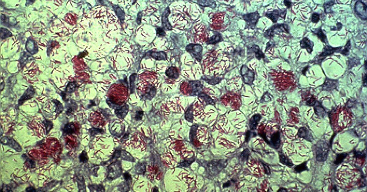
Table of contents
- Why the patch without sensation is a reasoning scenario
- The three cardinal signs
- How to test sensation and palpate nerves
- Slit-skin smear and biopsy: when they help
- Paucibacillary or multibacillary
- Treatment with multidrug therapy
- Leprosy reactions: type 1 and type 2
- Corticosteroids, nerves and follow-up
- Contacts, prophylaxis and stigma
- Outside the endemic zone: the delay in diagnosis
- How NEXO helps train this reasoning
- Frequently asked questions
- Final checklist
- References
At a basic health unit, a woman in her thirties comes in because of a light patch on her forearm that appeared a few months ago. She says it does not itch or hurt, and that she has already used two creams for ringworm without improvement. She also reports that, now and then, she feels tingling in the hand on the same side and that she burned herself on the stove without noticing. The first reaction of whoever attends her may be to think of yet another skin ringworm and change the cream. The scene is fictional and was written for teaching. This text uses this picture as a clinical reasoning scenario: why a patch without sensation changes everything, how to test sensation and palpate nerves, how to classify and treat leprosy and how to recognize the reactions that may appear before, during or after treatment.
NEXO is a daily medical diagnosis game: one clinical case a day, up to six progressive clues, and ICD-10 proximity feedback across five levels.
The text is not a treatment protocol. Regimens, doses, notification and follow-up follow national guidelines and the training of each service. The interest here is reasoning: why a simple exam, done with a piece of cotton, can decide the diagnosis and why worsening during treatment does not always mean failure. If you play the case of the day in NEXO, you will recognize the move: a clue that seems small can change the entire hypothesis.
Key points
- The diagnosis of leprosy is clinical and relies on at least one of three cardinal signs.
- The most useful sign in practice is the definite loss of sensation in a light or reddish patch.
- The classification into paucibacillary or multibacillary guides the duration of treatment with three drugs.
- Type 1 and type 2 reactions are inflammatory, can damage nerves and require prompt treatment, without suspending multidrug therapy.
- Examination of contacts and prophylaxis with a single dose of rifampicin help interrupt transmission.
Why the patch without sensation is a reasoning scenario
Leprosy is a chronic infection caused mainly by Mycobacterium leprae, which affects the skin and peripheral nerves. It is curable, but delay in diagnosis allows neural damage to advance and leave disabilities. Maymone and colleagues reviewed the clinical aspects and diagnostic techniques and recalled that the disease has a broad spectrum of presentations, which makes confusion with other skin diseases easier (Maymone and colleagues, 2020).
The 2018 World Health Organization guideline on diagnosis, treatment and prevention of leprosy is the reference used in this text (World Health Organization, 2018). It reinforces a central point: the diagnosis can be made by clinical examination, with or without laboratory tests.
What the scene teaches
The scene has three clues. The first is the patch that does not itch and does not respond to antifungals, which should already raise doubts about ringworm. The second is the tingling in the hand on the same side, which suggests nerve involvement. The third is the burn without pain, a sign of loss of sensation. Separated, each clue seems small. Together, they point to a disease that affects skin and nerve at the same time.
The post about differential diagnosis shows how small clues change the order of hypotheses.
The three cardinal signs
The 2018 World Health Organization guideline defines the diagnosis by the presence of at least one of three cardinal signs (World Health Organization, 2018):
- definite loss of sensation in a light or reddish patch of skin;
- thickened peripheral nerve, with loss of sensation or weakness in the muscles it innervates;
- acid-fast bacilli in the smear from skin slit scraping.
The first sign is the most accessible. A hypochromic or erythematous patch with clearly diminished sensation is very suggestive, especially in someone who lives or has lived in an area where the disease is common. The second sign requires training in palpation. The third depends on a laboratory, which is not always available.
A patch, yes, but which patch
Many diseases cause light patches: pityriasis versicolor, pityriasis alba, vitiligo, eczemas and scars. What sets leprosy apart is the loss of sensation inside the lesion. So, when faced with any light patch of slow evolution, testing sensation is a gesture worth making.
A spectrum, not a single disease
The presentation depends on each person's immune response. At one end, the tuberculoid form has few well-delimited lesions, with marked loss of sensation and few bacilli. At the other, the lepromatous form has many lesions, sometimes subtle and symmetrical, with many bacilli and diffuse nerve involvement. Between the two lie the borderline forms, more unstable and more prone to reactions. The indeterminate form, the initial one, may be just a light patch with mild change in sensation.
This spectrum explains why leprosy misleads. A single, well-delimited lesion can resemble a fungal infection or eczema. Multiple, infiltrated lesions can resemble other granulomatous diseases or skin lymphomas. In the lepromatous forms, loss of sensation in the lesions may be less evident, and nerve thickening gains weight.


How to test sensation and palpate nerves
The sensation test is simple, but it needs method. Explain what you are going to do, show the stimulus on a normal area and ask the person to close their eyes. Touch the lesion and the normal skin in varied order, with a cotton tip, and ask them to point to where they felt it. Always compare with the surrounding skin and with the opposite side.
Thermal and painful sensation are usually lost before tactile sensation. When resources are available, monofilaments of different calibers help grade the loss, especially on the hands and feet. A negative test on a small facial lesion may be less reliable, because the face has very rich sensation.
Palpate the nerves that matter
The nerves most often examined are the ulnar at the elbow, the median at the wrist, the superficial radial, the common fibular behind the head of the fibula, the posterior tibial behind the medial malleolus and the great auricular in the neck. Compare both sides, noting thickening, pain on palpation and consistency. Also test the strength of the muscles of the hands, of the feet and of eyelid closure.
In the scene, tingling in the hand suggests palpating the ulnar and median nerves and testing finger strength. Recording these findings at the start of treatment is important, because it allows any worsening to be noticed later. The post on signs, symptoms and history shows how the targeted exam completes what the person reports.
Eyes, hands and feet
The exam does not end at the skin and nerves. It is worth observing complete eyelid closure, corneal sensation when there is training for it and the presence of a red or dry eye. On the hands and feet, look for calluses, wounds, clawing of the fingers and areas of dry skin, signs that sensory and autonomic nerves have already been affected. The review by Gupta and colleagues recalls that neural damage is the main source of disability in leprosy (Gupta and colleagues, 2026).
These findings change the conversation with the person. Those who have lost sensation in their feet need to examine them every day, wear suitable footwear and seek the service when faced with any wound. Those who have difficulty closing their eyes need eye protection. The complete initial exam is the basis of this self-care routine and allows neural function to be compared at each follow-up.
Slit-skin smear and biopsy: when they help
The intradermal smear stained by the Ziehl-Neelsen method looks for acid-fast bacilli. When positive, it confirms the diagnosis and classifies the case as multibacillary. When negative, it does not rule out the disease, because many forms have few bacilli. The guideline recalls that the diagnosis remains clinical, with or without slit-skin smear and biopsy (World Health Organization, 2018).
Biopsy helps in doubtful cases, such as lesions without clear loss of sensation or conditions that imitate other diseases. The histological exam can show granulomas around nerves and skin appendages, as well as bacilli.
What the guideline does not recommend
The guideline does not recommend tests for latent infection in contacts. This means that, in practice, careful clinical examination of contacts remains the main tool for finding new cases. The post about pretest probability explains why a negative test carries different weight depending on the initial suspicion.


Paucibacillary or multibacillary
The operational classification in the 2018 World Health Organization guideline uses simple criteria, designed for services without a laboratory (World Health Organization, 2018). It determines the duration of treatment.
| Classification | Criteria | Treatment indicated in the guideline |
|---|---|---|
| Paucibacillary | One to five skin lesions, with no nerve involvement and a negative slit-skin smear | Rifampicin, dapsone and clofazimine for six months |
| Multibacillary | More than five lesions, or nerve involvement, or a positive slit-skin smear | Rifampicin, dapsone and clofazimine for twelve months |
| Rifampicin resistance | Confirmed by a specific test | Alternative twenty-four-month regimen, at a referral service |
When in doubt, multibacillary
When the classification is uncertain, the case is usually treated as multibacillary, because the risk of undertreating is greater than that of treating for longer. In the scene, the tingling in the hand suggests nerve involvement, which would already place the patient in the multibacillary group, even with a single patch.
Treatment with multidrug therapy
The 2018 guideline recommends the same three drugs for both forms: rifampicin, dapsone and clofazimine. The difference is in the duration, six months for the paucibacillary form and twelve for the multibacillary form (World Health Organization, 2018). Doses follow the national protocol and vary with age and weight.
Explaining the treatment is part of care. Clofazimine can darken the skin, which worries many patients and can lead to abandonment if it is not explained beforehand. Dapsone requires attention to anemia and hypersensitivity reactions. Rifampicin changes the color of urine. Knowing what to expect helps the person continue treatment to the end.
Following through to the end
Twelve months is a long time, and adherence can drop over the course of treatment, especially when the patches improve or when effects appear that the person did not expect. Combining regular follow-up visits, reviewing questions at each appointment and remembering that skin lesions can take time to disappear even with the infection cured helps prevent abandonment. Visual improvement is not the marker for the end of treatment: what defines discharge is completing the indicated regimen.
Curing the infection and protecting the nerve
Treatment eliminates the bacillus, but care does not end there. Damaged nerves may not recover, and the person needs to learn to protect hands, feet and eyes. Periodic examinations of nerve function, guidance on self-care and appropriate footwear are part of follow-up. The post about prognostic reasoning discusses how to think about what comes after the diagnosis.
Leprosy reactions: type 1 and type 2
Reactions are acute inflammatory episodes caused by the immune response to the bacillus. They can appear before the diagnosis, during treatment or after discharge. Gupta and colleagues reviewed the types, mechanisms and management (Gupta and colleagues, 2026).
Type 1 reaction, also called reversal reaction, appears mainly in borderline forms. Old lesions become redder, swollen and sometimes painful, and there may be neuritis, with pain along the nerve and loss of strength or sensation. It usually appears in the first months of treatment. Walker and Lockwood highlighted that type 1 reaction is an important cause of loss of nerve function, which can lead to disability and deformity, and that there is no consensus on the ideal dose or duration of the corticosteroid (Walker and Lockwood, 2008).
Type 2 reaction, or erythema nodosum leprosum, appears in lepromatous and borderline lepromatous forms. New, painful nodules appear, with fever, malaise and sometimes inflammation of the eyes, joints, testicles or nerves. The review by Gupta and colleagues notes that it must be distinguished from erythema nodosum of other causes, and also describes Lucio phenomenon, a rare and severe form.
Worsening is not treatment failure
A common pitfall is to interpret the reaction as failure of multidrug therapy or as allergy to the medicines. The review stresses that multidrug therapy should be started or maintained during the reaction (Gupta et al., 2026). Croskerry described how hasty interpretations lead to diagnostic errors (Croskerry, 2003). In leprosy, stopping treatment by mistake can prolong the disease and does not resolve the inflammation.
When the reaction is the first sign
The review by Gupta et al. recalls that, in many patients, the reaction is the first manifestation of leprosy (Gupta et al., 2026). A person may arrive with painful nodules and fever, or with skin lesions that suddenly swelled and severe pain in a nerve, without a prior diagnosis. In these cases, recognizing the reaction opens the way to diagnosing the underlying disease. The authors stress that identifying and treating early is what prevents deformities and disabilities.
Corticosteroids, nerves and follow-up
Corticosteroids are the basis of treatment for severe type 1 reactions and neuritis. The review by Gupta et al. cites the World Health Organization regimen with prednisolone in tapering doses over several weeks, and recalls the importance of rest and immobilization of the affected limb during neuritis. Doses follow the national protocol.
The evidence on long-term benefit is limited. A Cochrane review by Van Veen et al. found no significant difference in improvement of nerve function after twelve months between prednisolone and placebo, in trials with recent mild sensory loss or with nerve impairment of a few months to two years (Van Veen et al., 2016). This reinforces the importance of recognizing reactions early and monitoring nerve function closely.
Watch for adverse effects
Prolonged use of corticosteroids carries risks. Lun et al. reviewed the adverse effects in people with leprosy and found metabolic changes to be the most frequent, followed by infections, which accounted for most of the deaths associated with corticosteroids, especially tuberculosis (Lun et al., 2026). Screening for tuberculosis, parasitic infections and diabetes before and during use is part of care.
For type 2 reaction, thalidomide is an option in some countries, but it must not be used in women of childbearing age because of the risk to the fetus, as the review by Gupta et al. recalls. Clofazimine can also help in recurrent reactions.
Contacts, prophylaxis and stigma
People who live with someone who has leprosy have a higher risk of falling ill. Contact examination finds cases early, when there is still no disability. The 2018 guideline recommends a single dose of rifampicin for contacts from two years of age, after excluding leprosy and tuberculosis, and only with the patient's consent to reveal the diagnosis (World Health Organization, 2018). The same guideline recalls that BCG vaccine at birth has a protective effect.
The COLEP trial, by Moet et al., showed that a single dose of rifampicin reduced the occurrence of leprosy among close contacts by a little more than half in the first two years (Moet et al., 2008). The LPEP program, by Richardus et al., showed that the strategy is safe and feasible in routine services in several countries (Richardus et al., 2021).
How to examine a contact
Examining a contact follows the same script as the index case: inspection of all the skin in good light, sensitivity testing of any suspicious patch, palpation of the nerves and strength testing. Asking about numbness, tingling, painless wounds and weakness in the hands completes the assessment. When nothing is found, guidance on warning signs and an invitation to return if anything appears are part of care, because the disease can appear years later. The guideline also recalls that prophylaxis should only be implemented where contacts can be properly examined and followed up.
Consent protects against stigma
Leprosy still carries stigma in many places. That is why the guideline makes the approach to contacts conditional on the consent of the person who is ill. Discussing who will be informed, how and when is part of clinical reasoning as much as choosing the treatment regimen. The post on empathy and clinical reasoning discusses how listening helps in these conversations.
Outside the endemic zone: the delay in diagnosis
In places where leprosy is rare, diagnosis tends to be harder. Lockwood and colleagues described the patients seen at a national referral center in London over more than two decades. Most acquired the infection before migrating, symptoms appeared on average a few years after arrival, and there were cases in which diagnosis took up to twenty years. Dermatologists were the specialists who made the most diagnoses. The authors conclude that leprosy is not a disease of travelers, but of people who lived in an endemic area, and that clinicians need to recognize its signs (Lockwood and colleagues, 2022).
The long incubation period explains part of the problem. The disease can appear years after the person leaves the endemic area, and the question about where they lived is not always asked. A history of migration, a patch that does not improve and a vague neurological complaint form a combination that deserves attention anywhere.
The base rate changes the suspicion
The frequency of the disease in the place of origin changes the probability before the examination. The post on base rate in diagnosis shows how this data changes the reasoning, and the post on the diagnostic pause suggests a moment to ask what may be missing before changing the ointment once again.
How NEXO helps train this reasoning
In NEXO, the case of the day is fictional, with a real clinical presentation and up to six progressive clues. You propose a diagnosis by searching for the ICD-10 code and receive feedback on proximity in five levels, which shows whether the guess stayed at the exact code, the same category, the same group, the same chapter or a different chapter. This format speaks to the theme of this text: a neurological clue can change the chapter of a skin lesion.
To use the case of the day as training, try three habits. Before each guess, ask whether the skin lesion is related to any nerve. When the condition worsens during treatment, ask whether it is failure or reaction. After the result, note which clue would have anticipated the diagnosis.
Three exercises with cases
Test sensation mentally. Faced with a patch, ask what would change if it had no sensation.
Ask where the person comes from. List the diseases whose suspicion changes with a history of migration.
Separate worsening from failure. In cases with worsening during treatment, list the inflammatory explanations before concluding that the medication failed.
The post on learning based on clinical cases shows how cases help fix patterns that rarely appear in daily practice.
The limits of use of the game are described on the NEXO economy page. If you research teaching of clinical reasoning, the page for researchers explains how to get in touch. Professionals who write or review cases find information in curation and in creators, and institutions can learn about the proposal on the partnerships page.
Frequently asked questions
Can leprosy be diagnosed on clinical grounds alone?
Yes. The World Health Organization guideline accepts diagnosis based on the cardinal signs, with or without bacilloscopy and biopsy.
What is the most useful sign in practice?
The definite loss of sensation in a light or reddish patch, tested with a method and compared with normal skin.
Is leprosy curable?
Yes. Multidrug therapy with three drugs cures the infection, in six months for the paucibacillary form and in twelve for the multibacillary form.
Does multidrug therapy continue during a reaction?
Yes. The recommendation is to start or maintain treatment and treat the reaction at the same time.
Can contacts receive prophylaxis?
Yes. The guideline recommends a single dose of rifampicin for contacts from two years of age, after excluding leprosy and tuberculosis and with the patient's consent.
Can the disease appear years after leaving the endemic area?
Yes. The incubation period is long, so the history of where the person lived is always important.
Final checklist
- I tested the sensation of the patch and compared it with normal skin.
- I palpated the main nerves and tested strength of hands, feet and eyelids.
- I asked where the person was born and lived.
- I classified it as paucibacillary or multibacillary and, when in doubt, as multibacillary.
- I explained the expected effects of the three drugs.
- I gave guidance on signs of reaction and the need to return soon.
- I recorded neural function at the start and at each follow-up.
- I discussed the contacts with the patient's consent.
Want to practice your reasoning with progressive clues? Play the case of the day.
References
- Croskerry P. The importance of cognitive errors in diagnosis and strategies to minimize them. Acad Med. 2003. PMID 12915363.
- Gupta B, Singh M, Goldust M, et al. Reactions in leprosy: updated insights into pathophysiology, clinical spectrum, and therapeutic approaches. A narrative review. Health Sci Rep. 2026. PMID 42157916.
- Lockwood DN, McIntosh A, Armstrong M, et al. Diagnosing and treating leprosy in a non-endemic setting in a national centre, London, United Kingdom 1995-2018. PLoS Negl Trop Dis. 2022. PMID 36264976.
- Lun AI, de Barros B, Walker SL. A systematic review of adverse effects associated with systemic corticosteroids in the management of leprosy. PLoS Negl Trop Dis. 2026. PMID 41886473.
- Maymone MBC, Laughter M, Venkatesh S, et al. Leprosy: clinical aspects and diagnostic techniques. J Am Acad Dermatol. 2020. PMID 32229279.
- Moet FJ, Pahan D, Oskam L, Richardus JH. Effectiveness of single dose rifampicin in preventing leprosy in close contacts of patients with newly diagnosed leprosy: cluster randomised controlled trial. BMJ. 2008. PMID 18332051.
- World Health Organization. Guidelines for the diagnosis, treatment and prevention of leprosy. 2018. Document.
- Richardus JH, Tiwari A, Barth-Jaeggi T, et al. Leprosy post-exposure prophylaxis with single-dose rifampicin (LPEP): an international feasibility programme. Lancet Glob Health. 2021. PMID 33129378.
- Van Veen NH, Nicholls PG, Smith WC, Richardus JH. Corticosteroids for treating nerve damage in leprosy. Cochrane Database Syst Rev. 2016. PMID 27210895.
- Walker SL, Lockwood DN. Leprosy type 1 (reversal) reactions and their management. Lepr Rev. 2008. PMID 19274984.