GOLD 2026: one exacerbation changes COPD risk


Table of contents
- Why an exacerbation became a risk criterion
- What changed in groups A, B and E
- The new definition of exacerbation
- Severity by the Rome criteria
- When it is not COPD: the diagnoses that mimic exacerbation
- Cardiovascular risk after the crisis
- Initial treatment and follow-up
- Disease activity as a goal
- How NEXO helps train this reasoning
- Frequently asked questions
- Take-home messages
- References
A 68-year-old woman, a former smoker, with COPD diagnosed four years ago, returns for a routine visit. She reports that last winter she had a week of more cough, yellow sputum and shortness of breath greater than usual. She was seen at an urgent care unit, received prednisone for five days and an antibiotic, and improved without hospitalization. Today she is well, walks around the neighborhood and says the episode was "just a bad flu".
Under the old reading of the classification, a single episode treated outside the hospital left this patient in the same group as someone who had never had an attack. The 2026 edition of the Global Initiative for Chronic Obstructive Lung Disease report changed that calculation: a single moderate exacerbation is now enough to place the person in group E, the highest-risk group (GOLD, 2026). The "bad flu" has become a prognostic data point.
This text goes through what changed in GOLD 2026, focusing on exacerbation: why one attack counts so much, how it was redefined, how to measure severity without depending on where care was given, and which diseases masquerade as exacerbation in the emergency department.
Key points
- In GOLD 2026, a single moderate exacerbation in the past year already classifies the person in group E (GOLD, 2026).
- Exacerbation is now defined as worsening of dyspnea, cough or sputum that sets in within 14 days, with possible tachypnea or tachycardia.
- Severity is measured by the Rome criteria, with a scale of dyspnea, respiratory rate, heart rate, saturation, C-reactive protein and blood gas analysis.
- Pneumonia, pulmonary embolism, heart failure, myocardial infarction, arrhythmia and pneumothorax mimic exacerbation and must enter the differential.
- The risk of cardiovascular events rises sharply in the weeks after the attack and stays raised for months.
- The treatment goal is now low disease activity, with separate algorithms for initiation and follow-up.
Summary
- Why an exacerbation became a risk criterion
- What changed in groups A, B and E
- The new definition of exacerbation
- Severity by the Rome criteria
- When it is not COPD: the diagnoses that mimic exacerbation
- Cardiovascular risk after the attack
- Initial treatment and follow-up
- Disease activity as the goal
- How NEXO helps train this reasoning
- Frequently asked questions
- Take-home messages
Why an exacerbation became a risk criterion
COPD has a course marked by attacks. Each exacerbation accelerates the loss of lung function, worsens quality of life and increases the risk of death, and the best clue that someone will have an attack in the future is having had one in the past (GOLD, 2026). That was already in previous editions. What changed was the threshold.
Until the 2025 edition, group E required two or more moderate exacerbations in the previous year, or at least one that led to hospitalization. The executive summary of GOLD 2023, which created group E by merging the former groups C and D, already acknowledged that the number of attacks was the main driver of the classification (Agustí et al., 2023). In 2026, a single moderate attack became enough (GOLD, 2026).
The basis for this comes from studies that followed people from before diagnosis. A British cohort with primary care data showed that the annual rate of moderate exacerbations after diagnosis was 0.34 in those with no prior attack, 0.59 in those with one and 1.18 in those with two or more (Halpin et al., 2024). A single attack already nearly doubled the rate. In the same study, the eosinophil count added little to prediction once the history of attacks was known.
There is also a problem of label stability. In an analysis of three large clinical trials, the classification of "frequent exacerbator" changed in more than a third of patients from one year to the next, and the number of attacks in the previous year was a modest predictor of the following year (Sadatsafavi et al., 2021). If the label fluctuates so much, waiting for the second attack to act means leaving many people out of the group that benefits most from treatment adjustment.
The clinical reasoning behind the change is simple. The probability of a new attack depends on what has already happened, and a single moderate attack already shifts that probability in a relevant way, as we discussed in pretest probability. For the patient in the case, that means reviewing treatment now, not after the next attack.
What changed in groups A, B and E
The initial assessment remains in three steps: confirm the diagnosis by spirometry, grade the obstruction by forced expiratory volume in the first second and classify risk by symptoms and exacerbations (GOLD, 2026). Correct reading of the curve is still the starting point, and the text on spirometry details the choice between a fixed ratio and the lower limit of normal.
In the 2026 classification, group A brings together few symptoms and no moderate or severe exacerbation in the year. Group B brings together more symptoms, also with no crisis. Group E now includes those who had one or more moderate exacerbations, or at least one that led to hospitalization, regardless of symptoms (GOLD, 2026).
Symptoms continue to be measured by the modified Medical Research Council dyspnea scale and by the CAT questionnaire. The report recalls that these scales measure different things and that it is worth using the same tool throughout follow-up to compare the evolution (GOLD, 2026).
The table below summarizes the most practical changes for those who treat COPD.
| Change in GOLD 2026 | What changes in practice | Interpretation pitfall |
|---|---|---|
| One moderate exacerbation already defines group E | Review treatment after the first crisis treated outside the hospital | Considering the crisis "just a flu" and keeping the regimen |
| Definition with onset within 14 days | Worsening of dyspnea, cough and expectoration within a few days enters the definition | Calling a slow worsening over months an exacerbation |
| Severity by the Rome criteria | Measure dyspnea, vital signs, saturation, C-reactive protein and blood gas analysis | Classifying severity by the place of care |
| Explicit differential | Investigate pneumonia, embolism, heart failure and infarction | Closing the diagnosis of exacerbation by the history of COPD |
| Low disease activity as a goal | Absence of crises, of worsening symptoms and of accelerated loss of function | Confusing momentary stability with control |
| Separate algorithms for initiation and follow-up | Initial treatment by group and adjustment by response | Repeating the initial algorithm in those already treated |
| Oximetry and skin color | Suspect apparently normal saturation in dark skin | Ruling out hypoxemia based only on the oximeter |
The new definition of exacerbation
The previous definition spoke of acute worsening of respiratory symptoms that led to additional treatment. The problem with that formulation is that it depended on the decision of the person treating: the same picture could or could not be an exacerbation depending on whether the doctor prescribed corticosteroids or not.
GOLD 2026 adopts the definition built by the Rome proposal: an event characterized by dyspnea and or cough and expectoration that worsen within up to 14 days, which may be accompanied by tachypnea and or tachycardia, and which is usually associated with local and systemic inflammation caused by infection, pollution or another insult to the airways (GOLD, 2026).
This definition has three practical consequences. The first is time: a worsening that drags on for months calls for another explanation, such as disease progression, heart failure or depression. The second is the focus on symptoms, not on the prescription. The third is the reminder that the crisis has a cause, and looking for that cause is part of the assessment.
The most common triggers remain viral and bacterial infections, but air pollution enters the report explicitly (GOLD, 2026). On days of wildfire smoke, people with COPD are among the first to decompensate, as discussed in the text on who decompensates first.
The report also reinforces prevention. Vaccination against influenza, pneumococcus, covid-19, pertussis and respiratory syncytial virus has been updated, and the recommendation against the syncytial virus gained prominence for people with COPD (GOLD, 2026). The details of who is most at risk are in the text on respiratory syncytial virus. Smoking cessation remains the intervention that most changes the natural history of the disease, and brief advice fits into any consultation.
Severity by the Rome criteria
For a long time the severity of an exacerbation was measured by the place where it was treated. Mild was the attack treated only with a bronchodilator, moderate the one that required a corticosteroid or an antibiotic, severe the one that led to hospitalization. That classification depended on access to the service, on bed availability and on each team's habits.
The Rome criteria, incorporated into GOLD 2026, measure severity in the patient themself (GOLD, 2026). There are five variables: intensity of dyspnea on a visual analog scale from zero to ten, respiratory rate, heart rate, oxygen saturation and C-reactive protein. Arterial blood gas enters when available.
Mild attack
A mild attack is one in which dyspnea stays below 5 on the visual scale, respiratory rate below 24, heart rate below 95, resting saturation equal to or above 92 and with a variation of up to 3 points from the usual value, and C-reactive protein below 10 mg/L (GOLD, 2026).
Moderate attack
A moderate attack requires at least three of the five criteria: dyspnea equal to or above 5, respiratory rate equal to or above 24, heart rate equal to or above 95, saturation below 92 or a drop greater than 3 points, and C-reactive protein equal to or above 10 mg/L. If blood gas is available, the arterial oxygen pressure may be between 70 and 80 mmHg (GOLD, 2026).
Severe attack
A severe attack brings together the same clinical findings as the moderate one and is defined by blood gas: hypoxemia with arterial oxygen pressure equal to or below 60 mmHg and or hypercapnia above 45 mmHg with pH below 7.35 (GOLD, 2026). Respiratory acidosis is the dividing line, because it indicates the need for noninvasive ventilation and more intensive care.
When applied to hospitalized patients, the criteria show that the place of care is a poor indicator of severity. In a Dutch cohort of patients hospitalized for exacerbation, 14.3 in every 100 had a mild attack by the Rome criteria, 56.0 in every 100 had a moderate attack and 29.7 in every 100 had a severe attack (Reumkens et al., 2023). In-hospital mortality followed the classification, with 3.8, 6.9 and 13.9 deaths in every 100 in the mild, moderate and severe categories, which reinforces the prognostic value of the scale (Reumkens et al., 2023).
There is an important caution about saturation. Pulse oximeters overestimate saturation in people with dark skin, and the 2026 report includes this warning explicitly (GOLD, 2026). In a prospective study in critically ill patients with objective measurement of skin color, the difference between oximetry and blood gas increased in darker tones, with more occult hypoxemia (Hao et al., 2024). When in doubt, blood gas settles it.
When it is not COPD: the diagnoses that mimic exacerbation
Someone with COPD who arrives with more shortness of breath easily receives the label of exacerbation. It is the classic example of anchoring, discussed in the text on differential diagnosis: the prior history pulls the reasoning toward the most familiar hypothesis and ends the search too early.
GOLD 2026 lists the main diagnoses that should be considered: pneumonia, pulmonary embolism, heart failure, infarction, arrhythmias, pneumothorax and pleural effusion (GOLD, 2026). A good-practice review on the topic, signed by several authors of GOLD itself, proposed a structured assessment for the emergency department and recalled that a good part of readmissions after a hospitalization for exacerbation is due to other diseases, especially cardiovascular ones (Celli et al., 2023).
Pneumonia
Pneumonia is the most frequent differential. Chest radiography helps separate the pictures, and the presence of consolidation changes treatment and prognosis (Celli et al., 2023). High fever, pleuritic pain and focal findings on auscultation are clues in favor.
Pulmonary embolism
Embolism is harder to recognize, because dyspnea and tachycardia are common to both conditions. In a French multicenter study with patients hospitalized for acute respiratory worsening, embolism was confirmed in 5.9 in every 100 of the 740 participants, and more than half of the emboli were in main or lobar arteries (Couturaud et al., 2021).
This led to the logical question: is it worth looking for embolism in everyone? The SLICE trial, in Spain, randomized patients hospitalized for exacerbation to an active investigation strategy, with D-dimer and CT angiography when indicated, or to usual care. The active strategy did not improve the composite outcome of readmission, new embolism and death at 90 days (Jiménez et al., 2021). The message is to investigate based on clinical probability, as discussed in the text on pulmonary embolism, and to keep the suspicion when the picture does not fit, such as pleuritic pain without infection, worsening without sputum or venous thrombosis.
Heart failure and coronary syndrome
Decompensated heart failure can be indistinguishable from an exacerbation at the bedside. GOLD 2026 suggests considering natriuretic peptide and troponin in the assessment (GOLD, 2026). There is a detail: both can be raised in people with stable COPD, and the isolated value is less useful than the change from baseline and the clinical context (Celli et al., 2023).
The electrocardiogram and the echocardiogram complete the assessment when there is cardiac suspicion. People with COPD and heart failure with preserved ejection fraction are common, and the text on finerenone discusses this profile.
Cardiovascular risk after the crisis
One of the strongest messages of GOLD 2026 is that the exacerbation is also a cardiovascular event. The report describes an increased risk of infarction, arrhythmia, heart failure and stroke in the weeks following the crisis, with risk remaining raised for up to one year (GOLD, 2026).
A British cohort with more than 200 thousand people with COPD showed that, in the first 14 days after an exacerbation, the risk of a cardiovascular event was about three times higher than in periods without a crisis. After a severe exacerbation, this ratio reached 14.5 in the same interval, and the risk was still raised one year later (Graul et al., 2024).
The meta-analysis of the EXACOS-CV studies, with more than one million people in several countries, confirmed the pattern. The risk was highest in the first week after the crisis, with a hazard ratio near 10, and remained increased for about six months after a moderate crisis and for one year after a severe one (Nordon et al., 2025).
A Swedish cohort added a relevant finding for group E: even a single moderate exacerbation in the previous year was already associated with more risk of pulmonary embolism in follow-up, and the risk of infarction and embolism grew with the number and severity of crises (Wallström et al., 2024).
For practice, this has two implications. During the crisis, the electrocardiogram and troponin gain space when there is any suspicion. After the crisis, the follow-up visit is the moment to review blood pressure, lipids, antiplatelet therapy when indicated and adherence to cardiovascular treatment.
Initial treatment and follow-up
GOLD 2026 kept the logic of separating initial treatment, defined by the group, from follow-up treatment, defined by the response (GOLD, 2026). The change is in the weight that the first crisis gained in this account.
Initial treatment
For group E, the initial recommendation is the combination of a long-acting beta-agonist bronchodilator with a long-acting antimuscarinic. When blood eosinophils are at 300 cells per microliter or more, triple therapy with inhaled corticosteroid is considered (GOLD, 2026). The role of eosinophils and inhaled corticosteroid is detailed in the text on type 2 inflammation in COPD.
Follow-up
In follow-up, if the person keeps having crises with dual bronchodilation and eosinophils are at 100 or more, triple therapy is the suggested escalation. With eosinophils at 300 or more despite triple therapy, biologics become an option, and the text on dupilumab explores this point. Azithromycin, especially in former smokers, and roflumilast, in those with chronic bronchitis and more severe obstruction, remain as alternatives (GOLD, 2026).
During the crisis itself
In the treatment of the exacerbation, the report keeps the short-acting bronchodilator as the basis and systemic corticosteroid for a short period, equivalent to 40 mg of prednisone per day for five days. Eosinophils can guide the use of corticosteroid, and the report cites studies in which the eosinophil-guided strategy reduced exposure to corticosteroid without worsening the outcome (GOLD, 2026).
Antibiotics are indicated when there is increased sputum purulence associated with more dyspnea or a larger sputum volume, or when the person needs ventilation. The suggested duration in outpatients is up to five days. Point-of-care C-reactive protein can help reduce unnecessary prescriptions, while procalcitonin is not recommended for this decision (GOLD, 2026).
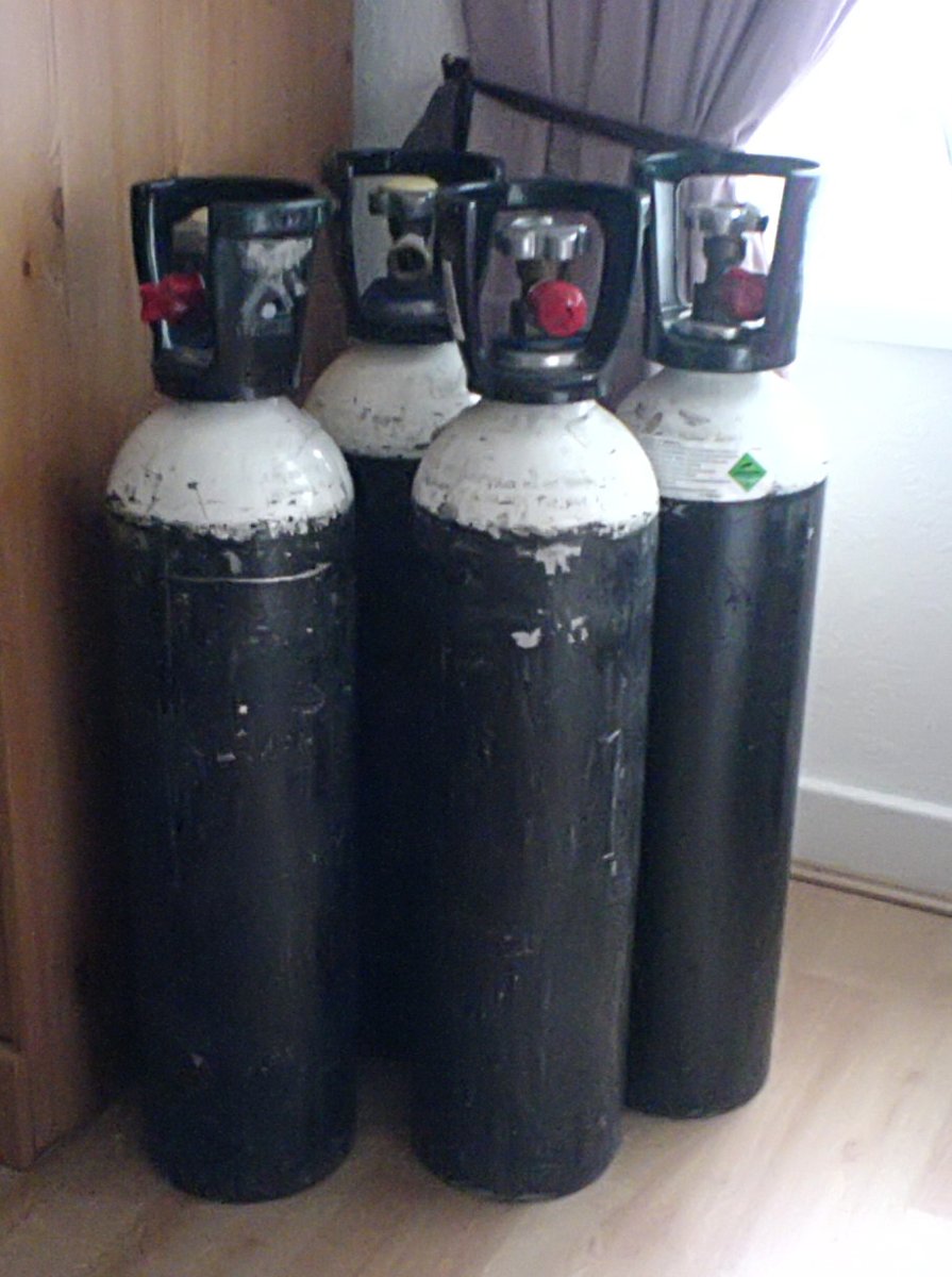
Oxygen during the exacerbation continues with a saturation target between 88 and 92, to reduce the risk of hypercapnia, and noninvasive ventilation is the first choice in respiratory acidosis (GOLD, 2026).
After discharge
The report recommends a review within one month after discharge from an admission for exacerbation. At this visit, it is appropriate to review inhaler technique, maintenance treatment, vaccination, referral to pulmonary rehabilitation and comorbidities, especially cardiovascular ones (GOLD, 2026).
Disease activity as a goal
The most important conceptual novelty of GOLD 2026 may be the treatment goal. The report proposes the concept of low disease activity, which combines absence of exacerbations, absence of worsening symptoms and absence of accelerated loss of lung function (GOLD, 2026).
The report separates two terms that are often confused. Stability describes the moment: the person is free of exacerbation and with symptoms at their usual pattern. Control describes the trajectory: over time, the disease is not progressing. Someone can be stable today and poorly controlled over the year, if they had two exacerbations and lost lung function faster than expected.
This separation brings COPD closer to the logic already used in asthma, in which the goal is to control the disease and reduce future risk, and not only to relieve the symptoms of the day. For the patient in the case, who feels well today, the reasoning is clear: today's stability coexists with a moderate exacerbation during the year, and the goal of low disease activity has not yet been reached.
The report also gained a new chapter on artificial intelligence and digital health, which discusses tools for reading spirometry, images and predicting exacerbations, with caution about biases and validation (GOLD, 2026). The key changes document also lists 14 new figures and the revision of the algorithms (GOLD, 2026, key changes).
How NEXO helps train this reasoning
In NEXO, the case of the day is fictional, with a real clinical presentation and up to six progressive clues, and you propose the diagnosis by searching for the ICD-10 code.
The limits of use of the game are described on the NEXO Economy page. If you research teaching of clinical reasoning, the page for Researchers explains how to get in touch. Professionals who write or review cases find information on Curator and on Creators, and institutions can learn about the proposal on the partnerships page. Searching for the right ICD-10 code completes this training.
Frequently asked questions
Does a single moderate exacerbation already place the person in group E?
Yes. In GOLD 2026, one moderate exacerbation in the last year, or one that led to admission, is enough to classify into group E, regardless of symptoms (GOLD, 2026).
What defines a moderate exacerbation?
According to the Rome criteria, a moderate exacerbation combines at least three of five findings: dyspnea equal to or above 5 on the visual scale, respiratory rate of 24 or more, heart rate of 95 or more, saturation below 92 or a drop greater than 3 points, and C-reactive protein of 10 mg/L or more.
Does being admitted mean the exacerbation is severe?
It depends on the findings. In a cohort of admitted patients, fewer than one third had a severe exacerbation by the Rome criteria, and most had a moderate exacerbation (Reumkens et al., 2023). Severe severity requires significant hypoxemia or hypercapnia with acidosis.
Should I look for pulmonary embolism in every exacerbation?
The investigation should follow clinical probability. Embolism appears in about 6 in every 100 patients admitted with respiratory worsening, but systematic screening in all of them did not improve outcomes in the SLICE trial (Jiménez et al., 2021).
Why order troponin and natriuretic peptide during the exacerbation?
Because heart failure and coronary syndrome mimic exacerbation, and cardiovascular risk rises sharply in the days after the crisis. The value must be read in context, since both can be raised in stable COPD (Celli et al., 2023).
Is the oximeter reliable for classifying severity?
It is useful, but it overestimates saturation in people with dark skin (Hao et al., 2024). When the clinical picture and the number diverge, arterial blood gas clarifies.
What is low disease activity?
It is the target proposed by GOLD 2026: no exacerbations, no worsening of symptoms and no accelerated loss of lung function over time. Stability describes the moment, and control describes that trajectory.
Take-home messages
- In GOLD 2026, a single moderate exacerbation already places the person in group E and calls for a treatment review.
- Exacerbation is defined by worsening of dyspnea, cough and sputum within 14 days, and it always has a cause to look for.
- Severity is measured in the patient, by the Rome criteria, and the oximeter can mislead on dark skin.
- Pneumonia, embolism, heart failure, infarction, arrhythmia and pneumothorax mimic the crisis and enter the differential.
- Cardiovascular risk is highest in the first weeks and remains raised for months, which makes follow-up within one month an opportunity for prevention.
- The goal is low disease activity: stability at the moment and control along the trajectory.
Want to train your reasoning when facing shortness of breath in someone who already has COPD? Play the case of the day on NEXO.
References
- Global Initiative for Chronic Obstructive Lung Disease. Global Strategy for the Diagnosis, Management, and Prevention of Chronic Obstructive Pulmonary Disease, 2026 Report. GOLD; 2026. GOLD 2026 Report.
- Global Initiative for Chronic Obstructive Lung Disease. GOLD 2026 Key Changes. GOLD; 2026. GOLD 2026 Key Changes.
- Agustí A, Celli BR, Criner GJ, et al. Global Initiative for Chronic Obstructive Lung Disease 2023 Report: GOLD Executive Summary. Eur Respir J. 2023. PMID 36858443. DOI 10.1183/13993003.00239-2023.
- Celli BR, Fabbri LM, Aaron SD, et al. Differential Diagnosis of Suspected Chronic Obstructive Pulmonary Disease Exacerbations in the Acute Care Setting: Best Practice. Am J Respir Crit Care Med. 2023. PMID 36701677. DOI 10.1164/rccm.202209-1795ci.
- Couturaud F, Bertoletti L, Pastre J, et al. Prevalence of Pulmonary Embolism Among Patients With COPD Hospitalized With Acutely Worsening Respiratory Symptoms. JAMA. 2021. PMID 33399840. DOI 10.1001/jama.2020.23567.
- Graul EL, Nordon C, Rhodes K, et al. Temporal Risk of Nonfatal Cardiovascular Events After Chronic Obstructive Pulmonary Disease Exacerbation: A Population-based Study. Am J Respir Crit Care Med. 2024. PMID 38127850. DOI 10.1164/rccm.202307-1122oc.
- Halpin DMG, Healey H, Skinner D, et al. Exacerbation history and blood eosinophil count prior to diagnosis of COPD and risk of subsequent exacerbations. Eur Respir J. 2024. PMID 39147410. DOI 10.1183/13993003.02240-2023.
- Hao S, Dempsey K, Matos J, et al. Utility of Skin Tone on Pulse Oximetry in Critically Ill Patients: A Prospective Cohort Study. Crit Care Explor. 2024. PMID 39268149. DOI 10.1097/cce.0000000000001133.
- Jiménez D, Agustí A, Tabernero E, et al. Effect of a Pulmonary Embolism Diagnostic Strategy on Clinical Outcomes in Patients Hospitalized for COPD Exacerbation: A Randomized Clinical Trial. JAMA. 2021. PMID 34609451. DOI 10.1001/jama.2021.14846.
- Nordon C, Simons SO, Marshall J, et al. The sustained increase of cardiovascular risk following COPD exacerbations: meta-analyses of the EXACOS-CV studies. ERJ Open Res. 2025. PMID 40524923. DOI 10.1183/23120541.01091-2024.
- Reumkens C, Endres A, Simons SO, et al. Application of the Rome severity classification of COPD exacerbations in a real-world cohort of hospitalised patients. ERJ Open Res. 2023. PMID 37228266. DOI 10.1183/23120541.00569-2022.
- Sadatsafavi M, McCormack J, Petkau J, et al. Should the number of acute exacerbations in the previous year be used to guide treatments in COPD? Eur Respir J. 2021. PMID 32855228. DOI 10.1183/13993003.02122-2020.
- Wallström O, Stridsman C, Lindberg A, et al. Exacerbation History and Risk of Myocardial Infarction and Pulmonary Embolism in COPD. Chest. 2024. PMID 39094732. DOI 10.1016/j.chest.2024.07.150.