Gastric cancer: staging gaps that cost a cure
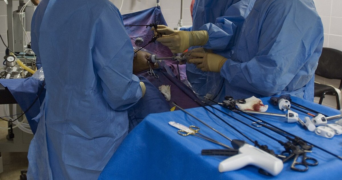
Table of contents
- Why this is a reasoning scenario
- The first clue: endoscopy and biopsy
- Imaging: what CT sees and what it misses
- Staging laparoscopy and peritoneal cytology
- After surgery: pathological staging and the plan
- Reasoning errors and the role of the team
- How NEXO helps train this reasoning
- Frequently asked questions
- Final checklist
- References
A 58-year-old woman comes to the outpatient clinic with early satiety, 6 kg of weight loss in three months and iron deficiency anemia. Endoscopy shows an ulcerated and infiltrative lesion in the gastric body, with thickened folds around it, and the biopsy confirms a poorly cohesive adenocarcinoma. CT of the chest, abdomen and pelvis describes 2 cm parietal thickening, a few perigastric lymph nodes of 9 mm and no liver lesions. The report ends with the phrase "no signs of distant disease". Surgery is scheduled for the following week, without endosonography, without laparoscopy and without going through the team meeting. On opening the abdomen, the surgeon finds small white nodules on the peritoneum and omentum. The scene is fictional and was written for teaching.
This text uses the case as a scenario for clinical reasoning for students and health professionals. The question is not which chemotherapy to use, but at what point along the way staging failed and what would have changed the decision. Every test has a known limit, and the error is almost never in a single test. It is in the sequence: a report read as a guarantee, a step skipped, a referral that did not happen. If you play the case of the day on NEXO, you will recognize the challenge of deciding with incomplete clues and knowing when a clue is still missing.
Key points
- CT is the basis of staging, but it sees small peritoneal disease poorly.
- Endosonography measures tumor depth and lymph nodes better than CT.
- Laparoscopy with peritoneal lavage finds metastases that no imaging test showed.
- Positive peritoneal cytology is already metastatic disease by staging and changes the plan.
- The number of lymph nodes examined in the specimen defines whether the pathological stage is reliable.
- The multidisciplinary meeting before the first treatment brings these pieces together in the right order.
Why this is a reasoning scenario
Locally advanced gastric cancer is a disease in which the first plan defines almost everything. If the disease is localized, the path tends to be perioperative chemotherapy and gastrectomy with lymphadenectomy. If there is already disease in the peritoneum, major surgery no longer brings benefit and brings only risk. A staging error, then, is not a detail of the report. It decides whether the patient receives treatment with curative intent or surgery that does not help.
What the scene teaches
The scene shows three common failures in sequence. The first is reading "no distant disease" as if CT had examined the peritoneum with the same precision with which it examines the liver. The second is skipping staging laparoscopy in an infiltrative type tumor, which is precisely the profile with the highest risk of peritoneal spread. The third is deciding on surgery without the team, and therefore without someone asking whether staging was complete.
What is not at stake
The text does not discuss the technique of gastrectomy, the choice of chemotherapy regimen or the treatment of peritoneal disease already established. These topics have their own posts in the oncology series. Here the focus is the earlier step: knowing what each test can and cannot show, and putting together the sequence that reduces the chance of a surprise in the operating room.
The first clue: endoscopy and biopsy
It all starts with endoscopy. It shows the location, the longitudinal extent and the macroscopic appearance of the tumor, and allows biopsies to be taken that are sufficient for diagnosis and for biomarkers. A small or single biopsy can give a correct diagnosis and still leave the case without material for the molecular tests that will guide systemic treatment.


The macroscopic type is a clue
The appearance of the lesion on endoscopy already signals risk. Diffuse infiltrative tumors and large ulcerated and infiltrative lesions carry a higher risk of peritoneal spread. The Asian adaptation of the ESMO guideline notes that Japanese experts, even without considering laparoscopy mandatory for everyone, recommend it for macroscopic type 4 or large type 3 disease, as the Japanese guideline provides (Shitara et al., 2024). In the scene, the thickened folds and the report of poorly cohesive carcinoma were warning signs that did not change the plan.
Biomarkers that need tissue
The pan-Asian guideline adapted from ESMO calls, at diagnosis, for assessment of HER2, microsatellite instability or mismatch repair deficiency, PD-L1 expression and, when available, claudin 18.2 by immunohistochemistry (Shitara et al., 2024). These results change systemic treatment, especially if the disease is metastatic. In practice, this requires several good-quality biopsies at the first endoscopy, so the exam does not have to be repeated weeks later.
- Take several biopsies from the edges and the center of the lesion.
- Record the exact location relative to the cardia and the pylorus.
- Describe the macroscopic type, because it signals peritoneal risk.
- Request the biomarkers at diagnosis, not only when the disease progresses.
- Mark the lesion when there is a chance of endoscopic treatment or minor surgery.
Imaging: what CT sees and what it misses
Contrast-enhanced CT of the chest, abdomen and pelvis is the basis of initial staging in all guidelines. The pan-Asian adaptation of ESMO recommends, at initial assessment, physical examination, complete blood count, liver and kidney function, upper gastrointestinal endoscopy and contrast-enhanced CT of the chest and the entire abdomen, including the pelvis (Shitara et al., 2024). The same text states that FDG PET is not recommended routinely.


The peritoneum is the blind spot
CT is good for liver and lung metastases and for enlarged distant lymph nodes. It is weak for small peritoneal implants, a few millimeters in size, which are the most common route of spread and carry the worst prognosis in gastric cancer (Burke, 2026). In a retrospective cohort of 95 patients assessed with CT, PET and laparoscopy, CT had 30 percent sensitivity for peritoneal disease in the group with localized disease, versus 85 percent for laparoscopy (Ofluoğlu et al., 2024).
Tumor depth is also misleading
CT also gets the depth of invasion wrong, and that depth defines the T stage. In a retrospective multicenter study of 191 operated patients, CT accuracy for T was 78 percent for T1, 55 percent for T2, 45 percent for T3 and 10 percent for T4 when compared with pathology (de Nucci et al., 2023). A T4 mistaken for T2 changes the indication for chemotherapy before surgery and the need for laparoscopy.
And PET?
FDG PET has a limited role in gastric cancer. Poorly cohesive and mucinous tumors take up little FDG, and small involved lymph nodes can go unnoticed. A review on occult lymph node metastasis describes that the maximum uptake value is affected by several factors and that new tracers are still under study (Ma et al., 2022). In the cohort of 95 patients, PET had 40 percent sensitivity for peritoneal disease in localized disease (Ofluoğlu et al., 2024).
Endoscopic ultrasound: measuring the wall and the lymph nodes
Endoscopic ultrasound shows the layers of the gastric wall and nearby lymph nodes with a resolution CT cannot reach. It is especially useful for separating early tumors, candidates for endoscopic resection or direct surgery, from tumors that have already gone beyond the muscularis propria and require treatment before surgery.
In the same multicenter study of 191 patients, linear endoscopic ultrasound had an accuracy of 100 percent in T1, 60 percent in T2, 74 percent in T3 and 80 percent in T4, and an accuracy of 85 percent for N, against 61 percent for computed tomography (de Nucci and colleagues, 2023). The biggest gain is precisely in T4 and N, where computed tomography errs most. Endoscopic ultrasound depends on the operator and on the tumor location, and large or stenosing lesions can prevent passage of the instrument.
A validation study used endoscopic ultrasound to separate patients into low risk (T1 or T2 without lymph node) and high risk (T3 or T4, or positive lymph node) before laparoscopy. Among 68 patients without metastasis on PET and computed tomography, laparoscopy found occult metastatic disease in 17, or 25 percent. None of the five low-risk patients had metastasis, and all cases of occult disease were in the high-risk group (Ayoub and colleagues, 2023). The authors themselves note that specificity was low and that the low-risk group was small.
Staging laparoscopy and peritoneal cytology
Staging laparoscopy is the examination that looks directly at the peritoneum. With it, the surgeon systematically inspects the peritoneal surface, the omentum, the liver and the pouch of Douglas, biopsies suspicious lesions and collects the wash for cytology. It is a short procedure, usually outpatient or with a brief hospital stay, and it is the step that most changes the plan in locally advanced tumors.


Who should undergo it
The pan-Asian adaptation of ESMO recommends laparoscopy and peritoneal wash for patients with resectable gastric cancer who are candidates for perioperative chemotherapy, and suggests considering them in selected patients going straight to surgery, with 100 percent panel consensus (Shitara and colleagues, 2024). A 2026 review describes Dutch and European Delphi consensus statements that defined when to perform laparoscopy and how to carry out the inspection and the wash (Burke, 2026). In the scene, the patient had a locally advanced tumor of the infiltrative type: she was a clear candidate.
What laparoscopy adds
In the cohort of 95 patients, laparoscopy had sensitivity and specificity of 92 and 80 percent in locally advanced disease, and of 95 and 99 percent in metastatic disease, while computed tomography had sensitivity of 16 percent and PET of 13 percent in the metastatic group (Ofluoğlu and colleagues, 2024). The study is retrospective, from a single center, and the numbers serve as an order of magnitude, not as fixed values.
Positive cytology is stage IV
Free tumor cells in the peritoneal wash, so-called positive cytology, classify the disease as metastatic even without visible implants. In a Japanese series of 89 patients with positive cytology and no other metastasis, operated on with macroscopically complete resection, five-year overall survival was 21.3 percent, and chemotherapy after surgery was one of the independent prognostic factors (Aoyama and colleagues, 2026). In the Turkish cohort, positive cytology was also associated with worse survival, with a hazard ratio of 1.80 (Ofluoğlu and colleagues, 2024).
The laboratory also needs a standard
Burke's review calls attention to a little-discussed point: a Japanese multicenter survey found more than tenfold variation in the rate of positive cytology among high-volume centers, linked to differences in laboratory method (Burke, 2026). If the same wash can be positive at one service and negative at another, the team needs to know the local method and discuss doubtful results with pathology.
- Inspect all quadrants of the peritoneum, including the pelvis and the diaphragm.
- Open the omental bursa when the tumor is on the posterior wall.
- Biopsy every suspicious lesion, even a small one.
- Collect the wash before manipulating the tumor.
- Send the material with a clear request for oncotic cytology.
- Record the finding in standardized language for the team meeting.
What each examination delivers
The table summarizes the role of each step and its main limitation. The numbers come from studies with different designs and should not be compared directly across rows.
| Step | Question it answers | Main limitation | Source |
|---|---|---|---|
| Endoscopy with biopsies | Diagnosis, location, macroscopic type, biomarkers | A small biopsy may not be enough for the biomarkers | Shitara 2024 |
| CT of the chest, abdomen and pelvis | Liver and lung metastases, distant lymph nodes | 30 percent sensitivity for the peritoneum in localized disease | Ofluoğlu 2024 |
| Endoscopic ultrasound | Depth (T) and lymph nodes (N) | Operator dependent; 60 percent accuracy for T2 | de Nucci 2023 |
| FDG PET | Distant metastasis in selected cases | Not routine; poorly cohesive tumors show low uptake | Shitara 2024 |
| Laparoscopy with lavage | Peritoneal implants and cytology | Invasive procedure; cytology varies between laboratories | Burke 2026 |
| Pathology of the specimen | Final pathological stage | Fewer lymph nodes examined reduce confidence in N | Zhao 2023 |
After surgery: pathological staging and the plan
Staging does not end in the operating room. Pathological N depends on the number of lymph nodes the surgeon removes and the pathologist examines. When few lymph nodes are examined, a patient with nodal disease may be classified at a lower stage, which removes the indication for adjuvant treatment. This phenomenon is called stage migration.
How many lymph nodes are enough
NCCN guidelines recommend examining at least 16 lymph nodes to reduce stage migration. A Chinese study of 12,670 gastrectomies found a U-shaped relationship between the number of lymph nodes examined and mortality, with an optimal threshold of 24 lymph nodes, and better survival in the group with 24 or more (Zhao et al., 2023). The same study showed that the average number of lymph nodes examined rose from about 18 to about 34 between 1999 and 2019, while the number of positive lymph nodes stayed stable.
What the team checks in the specimen
Before deciding on treatment after surgery, the team checks the number of lymph nodes examined, the status of the margins and, when there was prior treatment, the degree of tumor response. A report with few lymph nodes is not a reason to repeat the surgery, but it is a reason to read N with caution and discuss the case.
When staging changes treatment
The staging result separates very different paths. Japanese guidelines define when early gastric cancer can be treated with endoscopic resection alone, and the question is whether these criteria hold outside East Asia. At four centers in Australia, among 558 patients operated on without prior chemotherapy, 64 lesions met the Japanese 2021 absolute criteria for endoscopic resection, and 5 of them had positive lymph nodes at gastrectomy. The 55 lesions classified as eCura A or B had no positive lymph nodes (Young et al., 2025). The authors conclude that this endoscopic cure system could be applied safely in this Western population.
Locally advanced disease. In Western countries, perioperative chemotherapy with FLOT (fluorouracil, leucovorin, oxaliplatin and docetaxel) is the standard for locally advanced gastric adenocarcinoma, based on the FLOT4 and ESOPEC trials, while in East Asia surgery followed by adjuvant chemotherapy remains the most common standard (Okemoto et al., 2026). The same review describes the MATTERHORN trial, which showed a gain in event-free survival and overall survival by adding durvalumab to perioperative FLOT. For this patient, complete staging would have defined whether she was a candidate for this path.
Peritoneal disease. If laparoscopy had shown implants or positive cytology, the plan would be biomarker-guided systemic treatment, without gastrectomy at the outset. Strategies such as intraperitoneal chemotherapy and conversion surgery exist, but they are studied at selected centers, and Burke's review warns that variation in cytology can lead to wrong allocation in these treatments (Burke, 2026).
- Early disease confined to the mucosa: assess endoscopic treatment.
- Localized disease beyond the submucosa: surgery, with or without systemic treatment beforehand.
- Locally advanced disease: perioperative treatment according to the local guideline.
- Isolated positive cytology: discuss with the team, because the stage is already IV.
- Peritoneal implants: biomarker-guided systemic treatment.
Reasoning errors and the role of the team
Staging errors rarely come from a lack of knowledge. They come from shortcuts in reasoning, the same ones that appear in other areas of medicine. Knowing these shortcuts helps you recognize them in your own service.
Premature closure
A CT report with no metastasis gives the feeling that staging is finished. This is premature closure: accepting the first sufficient explanation and stopping the search. The post on the diagnostic pause shows how to create a fixed moment to ask what is still missing before deciding.
Overconfidence in the exam
Every exam has sensitivity and specificity, and a negative result on a low-sensitivity exam changes the probability little. Treating the report as absolute truth is a common bias. The post on how to choose exams and approaches discusses how the value of an exam depends on the question it answers.
Haste and schedule pressure
Patients with cancer want to start treatment soon, and so do teams. Haste leads to scheduling surgery before completing staging. A laparoscopy of a few days can avoid a laparotomy with no benefit and weeks of recovery before the right treatment.
The role of the multidisciplinary team
The multidisciplinary meeting brings together surgery, clinical oncology, radiology, pathology, gastroenterology, nutrition and nursing to decide the plan before the first treatment. There, staging is checked step by step, and the question "what is missing to decide?" is asked out loud. The post on interprofessional clinical reasoning shows why team decisions reduce individual errors.
Referral to a reference center
Gastrectomy with adequate lymphadenectomy, standardized staging laparoscopy and specialized reading of cytology depend on experience. When the service does not have this volume, early referral to a reference center is part of staging. Delay in referring also costs, and so the referral should go out together with the exam requests, not after them.
For the meeting, the team needs at hand the endoscopy report with macroscopic type and location, the biopsy and biomarker results, CT of the chest, abdomen and pelvis, endoscopic ultrasound when indicated, laparoscopy with lavage or the justification for not doing it, as well as nutritional status, organ function and the patient's preferences.
How NEXO helps train this reasoning
In NEXO, the case of the day is fictional, with a real clinical presentation and up to six progressive clues, and you propose the diagnosis by searching for the ICD-10 code. Staging of gastric cancer is a good exercise because each exam adds a clue and none of them closes the case alone. The post on differential diagnosis with clues shows how to weigh each finding, and the post on diagnostic uncertainty helps decide when doubt remains.
Three exercises with cases
Change the tumor. Say what changes if the lesion were a small, superficial 1.5 cm ulcer, with endoscopic ultrasound suggesting invasion of the mucosa only.
Change the result. Say what changes if laparoscopy showed no implants, but cytology came back positive.
Change the specimen. Say how you would read a gastrectomy report with N0 and only nine lymph nodes examined.
The limits of use of the game are described on the NEXO economy page. If you research teaching of clinical reasoning, the page for researchers explains how to get in touch. Professionals who write or review cases find information in curation and in creators, and institutions can learn about the proposal on the partnerships page.
Frequently asked questions
Does a normal CT rule out disease in the peritoneum?
No. CT sees small implants poorly, and in a cohort the sensitivity for peritoneal disease in localized disease was 30 percent (Ofluoğlu et al., 2024). That is why guidelines recommend laparoscopy in candidates for perioperative treatment.
Does every patient with gastric cancer need staging laparoscopy?
No. The pan-Asian guideline adapted from ESMO recommends it for candidates for perioperative chemotherapy and suggests considering it in selected patients going straight to surgery (Shitara et al., 2024). Early tumors rarely need it.
What does positive peritoneal cytology mean?
It means tumor cells free in the abdominal lavage. By staging, this is already metastatic disease, even without visible implants, and the plan should be discussed by the team.
Does PET replace laparoscopy?
No. FDG PET is not routinely recommended and has low sensitivity for peritoneal disease (Shitara et al., 2024; Ofluoğlu et al., 2024).
Why does the number of lymph nodes in the specimen matter?
Because few examined lymph nodes can hide nodal disease and lower the stage. The usual recommendation is to examine at least 16, and a large Chinese study suggested 24 as the optimal threshold (Zhao et al., 2023).
Final checklist
- I collected enough biopsies and requested the biomarkers at diagnosis.
- I recorded the macroscopic type and the tumor location.
- I requested contrast-enhanced tomography of the chest, abdomen and pelvis.
- I considered endoscopic ultrasound to define T and N.
- I indicated laparoscopy with lavage in locally advanced tumors.
- I took the case to the multidisciplinary meeting before the first treatment.
- I checked the number of lymph nodes and the margins in the specimen.
- I referred the patient early when the service did not have the volume for the case.
Want to practice your reasoning with progressive clues? Play the case of the day.
References
- Aoyama T, Cho H, Suematsu H, et al. Prognostic and Predictive Factors for Patients With Gastric Cancer who Are Positive for Peritoneal Lavage Cytology and Negative for Other Distant Metastasis. In Vivo. 2026. PMID 41760300.
- Ayoub F, Chapman CG, Chen H, et al. Endoscopic Ultrasound Predicts Risk of Occult Intra-Abdominal Metastases in Localized Gastric Cancer: A Validation Study. Gastroenterology Res. 2023. PMID 36895700.
- Burke E. Gastric Cancer Peritoneal Staging: Progress and Persistent Challenges. J Gastrointest Cancer. 2026. PMID 41634500.
- de Nucci G, Gabbani T, Impellizzeri G, et al. Linear EUS Accuracy in Preoperative Staging of Gastric Cancer: A Retrospective Multicenter Study. Diagnostics (Basel). 2023. PMID 37296694.
- Ma D, Zhang Y, Shao X, et al. PET/CT for Predicting Occult Lymph Node Metastasis in Gastric Cancer. Curr Oncol. 2022. PMID 36135082.
- Ofluoğlu CB, Aydın IC, Mülküt F, et al. Diagnostic Efficacy of Staging Laparoscopy Compared to CT and PET-CT in Gastric Cancer: A Retrospective Cohort Analysis. Medicina (Kaunas). 2024. PMID 39768958.
- Okemoto D, Nakayama I, Shitara K. Bridging paradigms and shaping the future of resectable gastric cancer: a narrative review of the post-MATTERHORN era. Gastroenterol Rep (Oxf). 2026. PMID 41809424.
- Shitara K, Fleitas T, Kawakami H, et al. Pan-Asian adapted ESMO Clinical Practice Guidelines for the diagnosis, treatment and follow-up of patients with gastric cancer. ESMO Open. 2024. PMID 38458658.
- Young E, Edwards L, Maurya A, et al. Lymph node metastases in early gastric cancer: Japanese Gastric Cancer Treatment Guidelines can be used for endoscopic resection in the West. Endosc Int Open. 2025. PMID 41403391.
- Zhao L, Zhang F, Jiao F, et al. The minimum number of examined lymph nodes was 24 for optimal survival of pathological T2-4 gastric cancer: a multi-center, hospital-based study covering 20 years of data. BMC Cancer. 2023. PMID 37735628.