Pneumothorax and flying: when is it safe to fly
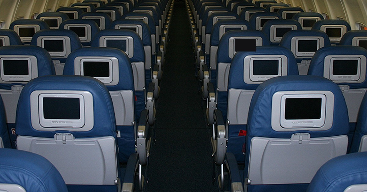
Table of contents
- Why this is a reasoning scenario
- The physics of the cabin: Boyle's law
- Who has primary spontaneous pneumothorax
- Pneumothorax, flying and diving
- The differential diagnosis on board
- Management on board
- On the ground: exams and the decision to treat
- When it is safe to fly again
- Who is at higher risk on a flight
- Reasoning: what remains from the case
- How NEXO helps train this reasoning
- Frequently asked questions
- Final checklist
- References
A 24-year-old man, tall and thin, a smoker, is on a long-haul night flight. Two hours after takeoff, he feels a sharp pain on the right side of his chest that worsens when he breathes deeply, and soon afterward he becomes short of breath. The crew asks a doctor among the passengers for help. The pulse oximeter in the onboard kit shows a lower saturation than expected for a healthy young man, and the cabin noise makes auscultation almost useless. The scene is fictional and was written for teaching.
This text uses the setting as a clinical reasoning scenario. The doctor must answer two questions at different times. The first is immediate: what may be happening and what to do with the resources on board. The second comes after landing: when this person can fly again. The text is not a summary of airline rules and gives no doses. If you play the case of the day on NEXO, you will recognize the challenge: the patient's profile weighs as much as the symptom.
Key points
- Sudden pleuritic pain with dyspnea in a tall young smoker suggests primary spontaneous pneumothorax.
- At cabin altitude, gas trapped in the chest expands according to Boyle's law.
- Auscultation on board is unreliable, and the differential includes pulmonary embolism and coronary syndrome.
- On the ground, the decision to treat depends on symptoms and high-risk signs, not only on size.
- The British recommendation is to wait 7 days after complete resolution on the radiograph before flying.
- Untreated pneumothorax is a contraindication to flying.
Why this is a reasoning scenario
The case brings together an environment with reduced pressure, limited resources and a common symptom. Chest pain on a flight can be muscular, anxiety-related, cardiac, pulmonary or pleural. The doctor on board has no radiograph or ultrasound, and the noise interferes with auscultation. They must decide using the history, inspection, pulse, respiratory rate and pulse oximeter.
The British thoracic society statement on air travel recalls a risk that seems unlikely and does happen: pneumothorax, pulmonary embolism and infarction can be mistaken for hyperventilation, with significant harm to the patient (Coker et al., 2022). In an anxious young man on a plane, the explanation of an anxiety attack appears early and closes the conversation too quickly.
What the scene teaches
The passenger brings clues that, together, change the probability. A young man, tall, thin and a smoker, with pain that worsens on inspiration and sudden-onset shortness of breath. The post on signs, symptoms and history shows how the profile of the person feeling the pain helps interpret the pain itself.
The physics of the cabin: Boyle's law
The cabin of a commercial aircraft is pressurized, but not to sea level. The British statement explains that, by Boyle's law, a cabin altitude of 2438 meters leads to an expansion of about 38 in every 100 of the volume of humidified gas (Coker et al., 2022). Gas trapped in a closed cavity, such as a pneumothorax or a pulmonary bulla, grows in the same proportion when the plane climbs.
The British summary for primary care describes the same effect: with altitude, the volume of air in cavities changes because pressure times volume remains constant, and a pneumothorax or a closed bulla expands and can cause respiratory discomfort (Josephs et al., 2013). The same cabin also reduces the pressure of inspired oxygen, and the two things add up in a partially collapsed lung.
What this means for the passenger
A small pneumothorax, which on the ground might be tolerated, can get bigger during the flight. This explains why the pain and dyspnea can worsen with cruise time and why saturation falls more than would be expected at sea level. The British statement places among those who need assessment before travel anyone who had a pneumothorax less than 6 weeks ago, those at higher risk, as in cystic lung disease or recurrent pneumothorax, and those with a trapped lung with chronic air space (Coker and colleagues, 2022).
Who has primary spontaneous pneumothorax
Primary spontaneous pneumothorax appears without known lung disease and is more common in young, tall men who smoke. The British statement notes that the risk of recurrence is higher in people with lung disease, in smokers and in tall men (Coker and colleagues, 2022). The passenger in the case has three of these characteristics.
In a Japanese hospital, among 938 episodes of spontaneous pneumothorax, about two in three were primary. In secondary cases, age and length of stay were greater, and smoking and surgery were less frequent (Onuki and colleagues, 2017).
The cause is usually hidden. In a CT study of healthy adults up to 40 years old, there were bullae or blebs in 7.2 per 100, and age, male sex and smoking were independent risk factors (Bresser and colleagues, 2024). A lung that looks normal can have small subpleural blebs that rupture.
Pathophysiology: the leak and the collapse
The normal pleural space has negative pressure, which keeps the lung expanded against the chest wall. When a subpleural bleb ruptures, air passes from the alveolus into the pleura, the negative pressure is lost and the lung partially collapses. The pain comes from irritation of the pleura, and the dyspnea comes from the loss of exchange area and the mismatch between ventilation and perfusion. The post on linking mechanism and symptom helps to use this kind of reasoning at the time of diagnosis.
In the cabin, the air already in the pleura expands, like any trapped gas. If the leak continues, the volume increases even more. The most severe form, tension pneumothorax, compresses the lung and the mediastinum and compromises venous return, with hypotension and instability.
Pneumothorax, flying and diving
The data on in-flight risk come mainly from diseases with lung cysts. In a questionnaire with patients with Birt-Hogg-Dubé syndrome, 13 of the 145 who had already traveled by plane had a spontaneous pneumothorax less than 1 month after a flight, and the estimated risk was 0.63 per 100 flights (Johannesma and colleagues, 2016). About one in five reported, after flying, symptoms that could be linked to pneumothorax.
Professional diving treats the lung with trapped air more strictly. In France, chest CT replaced radiography in the initial assessment of candidates for military diving starting in 2018. In a study with 307 candidates, air lesions such as cysts and bullae were much less frequent than benign nodules, but in general they led to disqualification (Bonnemaison and colleagues, 2022).
What these data teach
The authors of the Birt-Hogg-Dubé study suggest that symptoms during or soon after a flight may be the early phase of a pneumothorax, and they call for individual guidance (Johannesma and colleagues, 2016). For the passenger in the case, this means that an improvement on board does not end the investigation at the destination.
The differential diagnosis on board
On board, far from imaging, the doctor needs to keep several hypotheses open and look for signs that change management. The main hypotheses for sudden pleuritic pain with dyspnea on a flight are:
- Primary spontaneous pneumothorax.
- Pulmonary embolism, especially on a long flight with little mobility.
- Acute coronary syndrome, less likely in a young person, but serious.
- Pneumomediastinum, which can accompany pneumothorax.
- Musculoskeletal pain or viral pleuritis.
- Anxiety attack with hyperventilation, only after ruling out the rest.
Pulmonary embolism is the differential that worries most on a long flight, and the patient's profile helps little to separate it. Coronary syndrome is rare at 24 years old, but smoking does not exclude it. Anxiety comes last, because the British statement shows that labeling early as hyperventilation can cause harm (Coker and colleagues, 2022).
What to observe without a useful stethoscope
With auscultation compromised, inspection gains weight. Asymmetric chest expansion, high respiratory rate, tachycardia and falling saturation are signs the doctor can see and measure. Hypotension, tracheal deviation or rapid worsening point to tension pneumothorax, which is an emergency. The post on pretest probability shows how each sign changes the probability of each hypothesis, even without a confirmatory exam.
| Situation | What the source says | Source |
|---|---|---|
| Untreated closed pneumothorax | Most should not travel by plane | Coker and colleagues, 2022 |
| Resolved pneumothorax | Wait 7 days after complete resolution on the radiograph | Coker and colleagues, 2022 |
| Before the flight | Chest radiograph to confirm resolution | Josephs and colleagues, 2013 |
| Traumatic pneumothorax | 14-day wait in the British summary; limited evidence for 72 hours after drain removal | Josephs and colleagues, 2013; Coker and colleagues, 2022 |
| Chronic low-risk pneumothorax | Specialist assessment before the trip | Coker and colleagues, 2022 |
| After pleural surgery | Recurrence very unlikely | Josephs and colleagues, 2013 |
Management on board
On board, the options are few and the priorities are clear. Supplemental oxygen, a comfortable position, monitoring with an oximeter and respiratory rate, and conversation with the ground medical team the airline uses. If signs of instability appear, the discussion about diverting to the nearest airport enters the decision. The sources read do not define an oxygen flow for this situation, and so the text does not give that number.
Oxygen has a double rationale. It compensates for the reduced oxygen pressure in the cabin and helps with the hypoxemia caused by partial collapse. Decompression of a tension pneumothorax on board is an exceptional procedure, which depends on equipment, training and severity, and the sources read do not provide a protocol for it.
Record for those who receive
The doctor treating on board should record the start time, the course of saturation, the respiratory rate and what was done. This record helps the team receiving the passenger at the destination decide quickly. The post on clinical decision-making discusses how to choose the next step when resources are limited.


On the ground: exams and the decision to treat
After landing, the chest radiograph confirms the diagnosis in most cases, and CT comes in when there is doubt or to look for blebs. Cheng's review summarizes the recent change in guidelines: according to the 2023 British guideline, the patient's symptoms and the presence of high-risk features are the two most important factors in the initial treatment decision (Cheng and colleagues, 2024). The high-risk features cited are hemodynamic instability, significant hypoxia, bilateral pneumothorax, underlying lung disease, age over 50 with significant smoking and hemopneumothorax.
The same review describes that conservative management, without an invasive procedure, has become a main option for patients with primary pneumothorax who are stable and have few symptoms (Cheng and colleagues, 2024). In the RAMPP trial, with 236 patients, outpatient treatment with a device reduced the median hospital stay in the first 30 days from 4 days to 0, but had more adverse events (Hallifax and colleagues, 2020). For those with significant symptoms, aspiration or a drain remain options.
Thinking about recurrence
Recurrence is the problem that follows the patient after discharge. The review estimates recurrence at about 32 in every 100 in primary pneumothorax (Cheng et al., 2024). The authors describe that surgery, by thoracotomy or by video-assisted thoracoscopy, not only closes the leak but also prevents recurrence. The British summary for primary care states that recurrence is very unlikely after pleural surgery and that non-surgical pleurodesis is less effective (Josephs et al., 2013). A systematic review found recurrence between about 26 and 50 in every 100 with drainage alone, and from 0 to 3.2 in every 100 with talc applied during video-assisted thoracoscopy (Hallifax et al., 2017). In a cohort of 273 patients with primary pneumothorax, low BMI, large pneumothorax and non-surgical treatment were risk factors for recurrence (Tan et al., 2017).
Stopping smoking is part of the conversation. Smoking appears as a risk factor for blebs in the study by Bresser and for recurrence in the British statement (Bresser et al., 2024; Coker et al., 2022). For a 24-year-old man who travels a lot, this fact weighs on the decision between waiting and operating.
When it is safe to fly again
The question the passenger asks in the hospital is when they can board for the trip back. The 2022 British statement is direct: passengers should not travel by plane until 7 days after complete resolution on the chest radiograph (Coker et al., 2022). The same text says that most people with untreated closed pneumothorax should not travel and that those at higher risk of recurrence should be advised accordingly.
The British summary for primary care adds that, after a documented pneumothorax, a radiograph before the flight should confirm resolution, followed by a wait of 7 days for spontaneous pneumothorax or 14 days for traumatic pneumothorax (Josephs et al., 2013). The guidance for the passenger in this case therefore has two steps: confirm resolution on imaging and only then count the week.
Why the recommendations vary
The recommendations are not identical across sources. The 2013 British summary called for a follow-up radiograph and a wait of 7 days for spontaneous pneumothorax and 14 days for traumatic pneumothorax (Josephs et al., 2013). The 2022 statement speaks of 7 days after complete resolution and cites limited evidence that, in traumatic pneumothorax, flying 72 hours after drain removal, with the lung expanded, may be safe (Coker et al., 2022).
The British statement cites a study of 20 patients with small residual traumatic pneumothorax exposed to hypobaric hypoxia for 2 hours with no important clinical effect, but calls for caution because of the small number and the short exposure (Coker et al., 2022). For reasoning, the practical rule follows the most complete source, and the doubt is recorded.
Special situations
- Chronic low-risk pneumothorax: specialist assessment before travelling (Coker et al., 2022).
- Cystic lung disease: individual guidance because of the higher risk of recurrence.
- Traumatic pneumothorax: longer interval in the British summary.

Who is at higher risk on a flight
The question about risk does not apply only to those who have already had a pneumothorax. In the section on children, the British statement lists among those most exposed to the complications of reduced pressure those with trapped air inside the chest, such as recent pneumothorax or cystic lung disease (Coker et al., 2022). For all ages, the same text places pneumothorax of less than 6 weeks and recurrent pneumothorax among the situations that call for assessment before travel.
For those with underlying lung disease, the data are more reassuring than they seem. The British statement reports that the frequency of in-flight pneumothorax among passengers with lung disease appears to be low, and that in the British study of flight outcomes it was zero (Coker et al., 2022). The summary for primary care states that, in people with previous lung disease, flying does not appear to make pneumothorax more likely (Josephs et al., 2013). The greater risk lies in the gas that is already trapped, not in the flight itself.
Procedures that create air in the chest
Some procedures can leave a small pneumothorax that goes unnoticed. The British statement advises that, after interventional bronchoscopy with biopsy, those with pneumothorax on the follow-up radiograph should wait 1 week after resolution, and those without it should wait 1 week before flying (Coker et al., 2022). The same text notes that the risks of travel in these cases also include infection, pain and other symptoms that may require care at the destination.
After surgery, the type of procedure matters. The British statement describes that, after thoracotomy with surgical pleurodesis, there is no specific restriction on flying, and that reported recurrence after video-assisted thoracoscopy is higher (Coker et al., 2022). For the passenger in this case, who may need surgery if there is recurrence, this information is part of the conversation about the future.
Pneumomediastinum and other forms of barotrauma
Air that escapes from the alveolus does not always go only to the pleura. It can also travel to the mediastinum, and pneumomediastinum can accompany pneumothorax.
In the passenger's case, this differential changes little about management on board, but it changes what to look for on the ground. A radiograph that shows pneumothorax may also show air in the mediastinum, and CT helps when the picture is not explained by the pleura alone. The post on differential diagnosis with clues shows how to keep neighboring hypotheses open until the exam decides.
Reasoning: what remains from the case
The scenario teaches you to read the environment as part of the picture. Cabin pressure changes the volume of trapped gas and the availability of oxygen, and that changes the severity of a disease that would be calmer on the ground. The post on baseline rate in diagnosis notes that, in a tall young smoker, pneumothorax is a high-probability initial hypothesis.
The second lesson is about closing too early. An anxiety attack is an easy explanation on a plane, and the British statement shows that labeling early as hyperventilation can cause harm (Coker et al., 2022). The post on diagnostic pause helps build the habit of reviewing before concluding. The third lesson is about afterward: the diagnosis does not end at landing, and the decision to fly again is part of care, as discussed in the post on prognostic reasoning.
How NEXO helps train this reasoning
To use the case of the day as training, try three habits. Before the first guess, note the profile of the person with the symptom. With each clue, ask whether the environment changes the severity. After the result, see whether you labeled a benign explanation too early.
Three exercises with cases
Read the environment. For each case, write where the person was and how that place can change the disease.
List what you can measure. Without imaging, write the signs you can still observe and what each one changes.
Think about afterward. Write the question the patient will ask at discharge and what the source allows you to answer.
The post on case-based learning in clinical practice shows how cases help cement this habit.
The limits on use of the game are described on the NEXO economy page. If you research teaching of clinical reasoning, the page for researchers explains how to get in touch. Professionals who write or review cases find information under curation and creators, and institutions can learn about the proposal on the partnerships page.
Frequently asked questions
Does the gas in a pneumothorax expand in an airplane cabin?
Yes. By Boyle's law, a cabin altitude of 2438 meters expands the gas by about 38 in every 100 (Coker et al., 2022).
Can you fly after a resolved pneumothorax?
Yes. The British recommendation is to wait 7 days after complete resolution on chest radiography (Coker et al., 2022).
Is it worth having a radiograph before the flight?
Yes. The British summary calls for a radiograph before the flight to confirm resolution (Josephs et al., 2013).
Can a small, stable pneumothorax be treated without a drain?
Yes. Conservative management is an option for patients with stable primary pneumothorax and few symptoms (Cheng et al., 2024).
Does pleural surgery reduce recurrence?
Yes. After pleural surgery, recurrence is very unlikely (Josephs et al., 2013).
Final checklist
- I considered pneumothorax in sudden pleuritic pain with dyspnea.
- I took the profile into account: young, tall, thin and a smoker.
- I kept pulmonary embolism and coronary syndrome in the differential.
- I left the anxiety hypothesis for after ruling out the rest.
- I used inspection, respiratory rate, pulse and pulse oximeter when auscultation failed.
- I offered oxygen and spoke with the ground medical team.
- I looked for signs of tension pneumothorax.
- On the ground, I decided based on the symptom and the high-risk signs.
- I talked about recurrence, smoking and surgical options.
- I asked about recent thoracic procedures.
- I advised a follow-up X-ray and waiting 7 days after resolution before flying.
Want to practice your reasoning with progressive clues? Play the case of the day.
References
- Bonnemaison B, Castagna O, de Maistre S, et al. Chest CT scan for the screening of air anomalies at risk of pulmonary barotrauma for the initial medical assessment of fitness to dive in a military population. Front Physiol. 2022. PMID 36277200.
- Bresser MF, Wingelaar TT, Van Weering JAF, et al. An observational study ascertaining the prevalence of bullae and blebs in young, healthy adults and its possible implications for scuba diving. Front Physiol. 2024. PMID 38420621.
- Cheng HS, Wong C, Chiu PH, et al. Management of spontaneous pneumothorax: a mini-review on its latest evidence. J Thorac Dis. 2024. PMID 39144356.
- Coker RK, Armstrong A, Church AC, et al. BTS Clinical Statement on air travel for passengers with respiratory disease. Thorax. 2022. PMID 35228307.
- Hallifax RJ, McKeown E, Sivakumar P, et al. Ambulatory management of primary spontaneous pneumothorax: an open-label, randomised controlled trial. Lancet. 2020. PMID 32622394.
- Hallifax RJ, Yousuf A, Jones HE, et al. Effectiveness of chemical pleurodesis in spontaneous pneumothorax recurrence prevention: a systematic review. Thorax. 2017. PMID 27803156.
- Johannesma PC, van de Beek I, van der Wel JW, et al. Risk of spontaneous pneumothorax due to air travel and diving in patients with Birt-Hogg-Dubé syndrome. Springerplus. 2016. PMID 27652079.
- Josephs LK, Coker RK, Thomas M, et al. Managing patients with stable respiratory disease planning air travel: a primary care summary of the British Thoracic Society recommendations. Prim Care Respir J. 2013. PMID 23732637.
- Onuki T, Ueda S, Yamaoka M, et al. Primary and Secondary Spontaneous Pneumothorax: Prevalence, Clinical Features, and In-Hospital Mortality. Can Respir J. 2017. PMID 28386166.
- Tan J, Yang Y, Zhong J, et al. Association Between BMI and Recurrence of Primary Spontaneous Pneumothorax. World J Surg. 2017. PMID 27909771.