Laser in pilot's eyes: glare or retinal injury


 Alexei Peter dos Santos, Thiago Longo Moraes, and Caroline Wilhelmsen Martins
Table of contents
- Why a laser in the cockpit is a reasoning scenario
- When and where the laser strikes
- Functional effect: glare, flash blindness and afterimage
- Retinal injury: what is known
- Pathophysiology: how the laser damages the macula
- Symptoms that suggest injury
- The examination in the first days
- The differential diagnosis
- Management and follow-up
- Reasoning: what remains from the case
- How NEXO helps train this reasoning
- Frequently asked questions
- Take-home messages
- References
A 46-year-old captain was on final approach at night when a green beam came through the cockpit window. He reports that everything went white for a few seconds, that a spot remained in the center of his vision afterward, and that the instruments seemed dimmed. The first officer, who was not struck head-on, took over and landed. The next day, the captain seeks care: his eyes sting a little, the light bothers him, and he wants to know whether he "burned his retina." The scene is fictional and was written for teaching.
This text uses the case as a setting for clinical reasoning. The physician must answer two different questions. The first is functional: what the laser did to vision during the flight. The second is structural: whether there was retinal injury. The two call for distinct reasoning and distinct exams. The text is not a summary of aviation rules and does not give doses, because the sources reviewed do not define them for this scenario. If you play the case of the day on NEXO, you will recognize the challenge: a strong symptom does not always mean a serious injury, and a subtle injury can go unnoticed if the exam comes late.
Key points
- A laser in the cockpit usually causes a transient visual effect, such as glare and flash blindness.
- Retinal injury varies widely, and only the exam confirms or rules it out.
- Outside the cockpit, direct exposure to powerful handheld lasers can damage the macula.
- OCT shows findings typical of laser injury, some of them transient.
- The exam in the first days includes visual acuity, fundoscopy and OCT.
- The history of exposure is the clue that keeps the injury from being confused with other maculopathies.
Why a laser in the cockpit is a reasoning scenario
The case brings together a frightening event, strong symptoms and a question the patient asks in fear. The physician must separate the immediate effect on vision, which can disrupt the flight without leaving a mark, from tissue injury, which can leave sequelae. Confusing the two leads to two opposite errors: reassuring someone who has an injury or alarming someone who does not.
What the scene teaches
The captain brings clues of two kinds. The white spot, the afterimage and the difficulty reading the instruments are functional effects. The stinging and light sensitivity the next day are complaints that call for an exam. The scenario trains you not to let the fright of the account decide severity on its own. The post on signs, symptoms and history shows how to separate what the person felt from what the exam can confirm.
When and where the laser strikes
Most laser illuminations happen in phases of flight when the pilot most needs vision. Between 2004 and 2008, the proportion of illuminations at about 610 meters of altitude or less rose from 12.5 to 26.7 per 100 (Nakagawara and colleagues, report, 2010). In the same period, about 70 per 100 of the events with a recorded time occurred between 19:00 and 23:00 (Nakagawara and colleagues, report, 2011). Takeoff and landing, at night, are precisely the phases with the least margin for error.
The combination of darkness, an eye adapted to low light and a very bright source explains why the effect is so strong.
Functional effect: glare, flash blindness and afterimage
The technical report from the American medical institute defines three transient visual effects (Nakagawara and colleagues, report, 2004). Glare is the loss of vision of an object because a strong light is near the same line of sight. Flash blindness is an interference that persists after the light goes out. The afterimage is a transient image that remains in the visual field after exposure.
The same report explains that when someone adapting to half-light is exposed to very intense light, temporary visual impairment is almost certain, and that the effects can last from seconds to minutes, while full dark adaptation can take 30 minutes or more (Nakagawara et al., report, 2004). In this case, that explains why the instruments seemed to go dark even after the beam had left the window.
What the simulator showed
In the simulator study with 34 pilots, the most reported effects were glare in 30.9 and flash blindness in 30.9 per 100 responses, followed by afterimage in 13.0, and in 25.2 per 100 responses there was no effect (Nakagawara et al., report, 2004). Multiple and more severe effects increased as exposure increased.
The report also cites reference levels used by the agency for airspace near airports: about 100 µW/cm² as the level at which flash blindness and afterimage can hinder the pilot, and 5 µW/cm² as the level at which glare can be significant (Nakagawara et al., report, 2004). These levels are far below those capable of damaging tissue, which shows why the functional effect can exist without injury.

Retinal injury: what is known
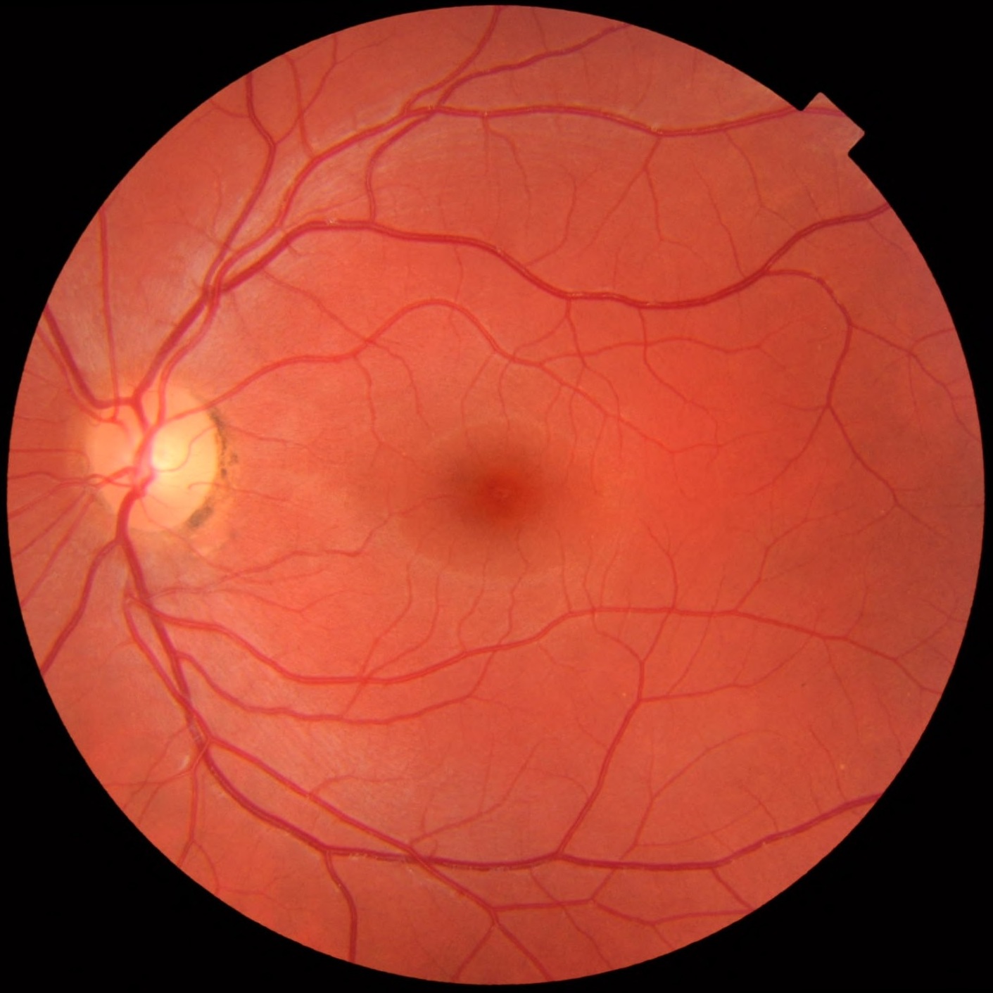
The commander's question, whether it "burned the retina", has no answer before the examination. The sources read in full do not include a series of pilots examined after a laser in the cockpit, and so this text does not estimate the chance of injury in that group. What they show is that the presentation varies greatly depending on the type of laser, the exposure time and the way the beam hit the eye (Neffendorf et al., 2019).
Why the answer is not always "no"
Outside the cockpit, the literature on handheld lasers shows real injuries. The Oxford review describes sequelae ranging from severe loss of acuity to no evident clinical impact: mild injuries tend to affect only one eye and can resemble a macular dystrophy, while severe ones can leave large atrophic scars on the retina (Neffendorf et al., 2019). Clinical reasoning, therefore, does not start from the conclusion that there is no injury. It starts from the examination.
Pathophysiology: how the laser damages the macula
The swept-source OCT study describes the mechanism in two parts. The retinal pigment epithelium has a lot of melanin, which absorbs the laser energy and converts it into heat, causing photothermal injury with protein denaturation, cell rupture and secondary inflammation (Moussa et al., 2025). In addition, there is photochemical injury, with production of free radicals and programmed death of pigment epithelium cells and photoreceptors.
The same authors explain that short-wavelength lasers, such as blue, have more energy, penetrate less and are absorbed preferentially by melanin, which makes them more harmful (Moussa et al., 2025). Heat dissipation up to the layer of Henle fibers causes opacification and thickening, which appear as hyperreflective bands on OCT. Focal rupture of Bruch's membrane can open the way for secondary neovascularization.
Blinking does not protect
A case report recalls that, with class 3 lasers above 5 mW, injury can occur in an instant and that blinking or looking away does not protect (Gregori et al., 2024). The same text warns that device labels can be inaccurate. In another case, a pointer labeled below 5 mW had a measured power about twenty times higher (Dhrami-Gavazi et al., 2015). For the physician, this means the stated power cannot be used to rule out injury.


Symptoms that suggest injury
The functional effect passes in seconds or minutes. When the symptom persists, the question changes. In Moussa's series, the symptom most reported by those with laser macular injury was central scotoma, in 61 out of every 100 eyes, and the rest reported blurred vision (Moussa et al., 2025). More than half of the eyes had acuity worse than 20/40 at the first visit.
In the commander's case, the spot in the center of vision during the flight is consistent with an afterimage, which is temporary. What would matter the next day is knowing whether that spot is still there. A fixed spot in the center, distortion of lines or a drop in acuity are the complaints that shift the reasoning from functional effect to structural injury.
The history that changes the case
The review on injuries in children notes that the presentation depends on the type of laser, the exposure time and the way the beam reached the eye (Neffendorf et al., 2019). The history therefore needs to cover these variables. The useful questions are the color of the beam, whether the gaze was direct or from the side, how long it lasted, whether there was more than one exposure and whether one or both eyes were hit.
The shape of the injury also tells a story. In Gregori's report, curvilinear streak lesions in the macula pointed to self-inflicted injury, while injuries caused by another person tend to be focal in the fovea (Gregori et al., 2024). The same text notes that a beam reflected off a mirrored surface can act as a direct beam. In Moussa's series, small satellite lesions around the main one indicated repeated exposure (Moussa et al., 2025).
Why age weighs on the prognosis
The patients in Moussa's series were mostly children and adolescents, and they had a relatively good visual outcome despite the dramatic images (Moussa et al., 2025). The authors attribute this in part to the healthy Bruch's membrane of young people, which helps the pigment epithelium and retina heal. An adult pilot is not in this group, and the sources read do not allow estimating his prognosis from these data.
In practice, this means the reasoning should be based on the patient's own examination, not on the average of a published series. If the fundus photograph, the OCT and the acuity are normal, that is the strongest finding, and it is worth more than any estimate taken from another population. If there is an abnormality, follow-up with a retina specialist defines what happens next.
It is also worth recording in the chart the date and time of the event, the phase of the flight, the color of the beam and the symptoms described by the pilot in his own words. This record helps compare exams over time and serves the notification that many authorities request. A detailed account made on the same day is usually more reliable than memory reconstructed weeks later, when the pilot has already heard versions of the case from colleagues and the press.
The examination in the first days
The Oxford review says the diagnosis calls for a targeted history and high-quality methods: indirect ophthalmoscopy and OCT for diagnosis and follow-up, with infrared fundus photography and fundus autofluorescence as support (Neffendorf et al., 2019). Imaging answers the question about structure. The question about function calls for measuring acuity with proper correction, and the post does not detail other tests because the sources read do not provide a protocol for pilots.
In a series of 17 consecutive patients with injury from recreational laser pointers, all eyes underwent a complete examination of the anterior and posterior segments and spectral-domain OCT, with reassessment of acuity and OCT at one and three months (Joshi et al., 2025). OCT showed defects of the outer retinal layers, lamellar holes, hemorrhages at different levels and a full-thickness macular hole.
Time matters. In the report of a young man who looked directly at a red pointer, the curvilinear hyperreflective bands that followed the fibers of Henle appeared on OCT soon after the injury and had largely disappeared within two weeks (Dhrami-Gavazi et al., 2015). The authors conclude that this finding is highly specific to laser injury but can vanish within a few days. A late examination may miss the most characteristic clue.
What OCT can show
Moussa's study describes the acute and chronic findings (Moussa et al., 2025). The acute ones include focal or diffuse disruption of the outer retinal layers, a hyperreflective subretinal elevation, the anvil-shaped lesion and the angular sign of hyperreflectivity of the Henle fiber layer. The chronic ones include secondary choroidal neovascularization and macular hole, which may be full-thickness or lamellar, with characteristic straight edges.
| Finding | What it suggests | Source |
|---|---|---|
| Glare, flash blindness or afterimage during flight | Transient functional effect | Nakagawara, report, 2004 |
| Curvilinear hyperreflective bands in the Henle fiber layer | Acute laser injury, a finding that may disappear within weeks | Dhrami-Gavazi et al., 2015 |
| Defect or hole in the outer retinal layers | Structural lesion of the macula | Moussa et al., 2025 |
| Full-thickness macular hole | Severe lesion, which may appear weeks later | Gregori et al., 2024 |
| Secondary choroidal neovascularization | Complication from rupture of Bruch's membrane | Moussa et al., 2025 |
The differential diagnosis
When the patient reports the exposure, the diagnosis seems obvious. The challenge appears when the history does not come. A review of injuries in children warns that laser retinopathy can be mistaken for retinal dystrophies, especially if the exposure is neither reported nor asked about (Neffendorf et al., 2019). In Moussa's study, the exclusion criteria show the conditions the authors had to rule out before attributing the lesion to the laser (Moussa et al., 2025).
- Macular dystrophies.
- Maculopathy after ocular trauma.
- Uveitis and intraocular infection.
- Retinal vascular disease.
- Myopic maculopathy.
- Drug-induced maculopathy.
Closing too early
In the commander's case, there are two risks of ending the reasoning too soon. One is concluding that "it was just the scare" without examining. The other is concluding that "the retina was burned" from the intensity of the report. Croskerry describes this error as the tendency to accept the first plausible diagnosis without checking (Croskerry, 2003). The post on differential diagnosis with clues shows how to keep the hypotheses open until the examination decides.
Management and follow-up
If the commander's examination is normal, with visual acuity, fundoscopy and OCT showing no changes, that examination is the strongest finding. The sources read do not provide a follow-up protocol for pilots without lesions, and so the text does not define timeframes. The return to flight follows each country's rules and the examiner's assessment, which this text does not summarize.
If there is a lesion, imaging follow-up is part of the treatment. Gregori's case report describes that spectral-domain OCT is sensitive for assessing the lesion and tracking its course (Gregori et al., 2024). In the same case, blue laser light reflected in a window caused a full-thickness macular hole in both eyes after one month.
Macular hole and neovascularization
Laser-induced macular holes appear among the chronic findings in Moussa's series, with characteristic straight edges (Moussa et al., 2025). A case report published in 2026 summarizes that there is no consensus on treatment: the approaches described range from observation to topical, systemic or intravitreal corticosteroids, and antiangiogenic agents when there is choroidal neovascularization, with no solid evidence of benefit (Ouacha and De Temmerman, 2026). The authors say the prognosis seems to depend more on the severity of the initial lesion and the recovery potential of the fovea; in the case described, visual acuity improved even with persistent outer retinal changes on OCT. The decision rests with the retina specialist. In Moussa's series, secondary neovascularization regressed with intravitreal antiangiogenic injections (Moussa et al., 2025). The sources read do not provide a regimen that applies to every case, and so this text does not include doses.


A possible conversation with the commander
- Explain the difference between a transient effect and a retinal lesion.
- Say that the complete examination answers the question better than the report.
- Perform OCT examination in the first days, when the typical findings still appear.
- Arrange a follow-up visit if a fixed spot, distortion or vision loss appears.
- Record the event for follow-up and for the flight assessment.
Reasoning: what remains from the case
The scenario teaches you to separate two questions that the patient merges into one. "Did the laser interfere with my vision?" is a question about function, and the answer is almost always yes. "Did the laser damage my eye?" is a question about structure, and the answer depends on the examination. The doctor who answers the second based on the first errs in one direction or the other.
The second lesson is about time. Some OCT findings last only a few days, and a late examination may not find them (Dhrami-Gavazi et al., 2015). The third is about history: without the right question, a laser injury can be labeled as another disease. The post on diagnostic pause helps build the habit of reviewing before concluding, and the text on base rate in diagnosis reminds us that the rarity of the injury in pilots is a data point, not a guarantee.
How NEXO helps train this reasoning
To use the case of the day as training, try three habits. Before the first guess, separate the function clues from the structure clues. With each clue, ask whether it confirms or only alarms. After the result, note whether you decided based on the intensity of the account.
Three exercises with cases
Separate function and structure. For each case, write down what the person felt and what an examination could show.
Think about time. Ask whether any finding could disappear before the examination and when would be the best time to examine.
Look for the missing history. Write down which exposure or event would change the diagnosis if the person mentioned it.
The post on learning based on clinical cases shows how cases help cement this habit.
The limits on using the game are described on the NEXO Economy page. If you research the teaching of clinical reasoning, the page for Researchers explains how to get in touch. Professionals who write or review cases find information on Curator and on Creators, and institutions can learn about the proposal on the partnerships page.
Frequently asked questions
Can a laser in the cockpit interfere with a pilot's vision?
Yes. Glare, flash blindness and afterimage were the most reported effects in a simulator (Nakagawara et al., report, 2004).
Does a laser injury always look the same?
No. The presentation depends on the type of laser, the exposure time and how the beam hit the eye (Neffendorf et al., 2019).
Does OCT help recognize a laser injury?
Yes. It shows characteristic findings in the acute and chronic phases (Moussa et al., 2025).
Is it worth examining in the first few days?
Yes. Some typical OCT findings can disappear in about two weeks (Dhrami-Gavazi et al., 2015).
Can the injury worsen after the event?
Yes. In a case report, blue laser light reflected off a window caused a full-thickness macular hole in both eyes after one month (Gregori et al., 2024).
Take-home messages
- I separated the functional effect during the flight from the possibility of injury.
- I asked about the beam color, the duration and the phase of the flight.
- I measured visual acuity with proper correction.
- I performed indirect ophthalmoscopy and considered infrared and autofluorescence.
- I requested OCT in the first days after exposure.
- I considered the differential with dystrophies and other maculopathies.
- I arranged a follow-up visit if a fixed spot, distortion or vision loss appears.
- I planned imaging follow-up when there was an injury.
- I remembered that the rules for returning to flight vary between countries.
Want to practice reasoning with progressive clues? Play the case of the day.
References
- Croskerry P. The importance of cognitive errors in diagnosis and strategies to minimize them. Acad Med. 2003. PMID 12915363.
- Dhrami-Gavazi E, Lee W, Balaratnasingam C, et al. Multimodal imaging documentation of rapid evolution of retinal changes in handheld laser-induced maculopathy. Int J Retina Vitreous. 2015. PMID 27847607.
- Gregori NZ, Cai L, Moshiri Y. Self-Inflicted Laser-Induced Retinopathy. Diagnostics (Basel). 2024. PMID 38396400.
- Joshi SM, Maanju S, Vibhute G, et al. Clinical and Imaging Spectrum of Laser-Induced Maculopathy Caused by Laser Projection Devices. Cureus. 2025. PMID 41246636.
- Moussa M, Leila M, Elashri MF, et al. A swept-source optical coherence tomography study of the spectrum of laser pointer maculopathy. Int J Retina Vitreous. 2025. PMID 40474312.
- Nakagawara VB, Montgomery RW, Dillard AE, et al. The effects of laser illumination on operational and visual performance of pilots during final approach. Federal Aviation Administration, Office of Aerospace Medicine, DOT/FAA/AM-04/9. 2004. Technical report.
- Nakagawara VB, Montgomery RW, Wood KJ. Laser Illumination of Flight Crew Personnel by Month, Day of Week, and Time of Day for a 5-Year Study Period: 2004-2008. Federal Aviation Administration, Office of Aerospace Medicine, DOT/FAA/AM-11/7. 2011. Technical report.
- Nakagawara VB, Montgomery RW, Wood KJ. The Illumination of Aircraft at Altitude by Laser Beams: A 5-Year Study Period (2004-2008). Federal Aviation Administration, Office of Aerospace Medicine, DOT/FAA/AM-10/21. 2010. Technical report.
- Neffendorf JE, Hildebrand GD, Downes SM. Handheld laser devices and laser-induced retinopathy (LIR) in children: an overview of the literature. Eye (Lond). 2019. PMID 30894692.
- Ouacha MA, De Temmerman S. Holiday Souvenir Gone Wrong: Laser Pointer-Induced Maculopathy. Cureus. 2026. PMID 42367396.