Ectopic pregnancy: beta-hCG, ultrasound and doubt

Table of contents
- Why ectopic is a reasoning problem
- Pregnancy of unknown location
- Transvaginal ultrasound as the central exam
- Beta-hCG: from a single value to the curve
- Risk models: who needs intensive follow-up
- Instability and step-by-step reasoning
- How NEXO helps train this reasoning
- Frequently asked questions
- Take-home messages
- References
A 29-year-old woman arrives at the emergency department with mild vaginal bleeding and slight lower abdominal pain. The pregnancy test is positive and she is not sure of the date of her last menstrual period. Transvaginal ultrasound shows an empty uterus and no adnexal mass. The beta-hCG comes back at an intermediate value. The team's question is direct: is this an early normal pregnancy, an ongoing miscarriage or an ectopic pregnancy?
This scene is common and uncomfortable. The test does not answer right away, the patient wants to know what is happening and the team knows that an unrecognized ectopic can rupture. At the same time, acting too fast can interrupt a desired, viable pregnancy.
This text shows how to reason in this scenario: what pregnancy of unknown location means, why the discriminatory zone has lost strength, how to read the beta-hCG curve, what ultrasound shows and how risk models help decide who needs closer follow-up.
Key points
- Transvaginal ultrasound is the central test, and beta-hCG helps interpret what it shows.
- An empty uterus with a positive test is a pregnancy of unknown location, not a diagnosis of ectopic.
- No single beta-hCG value safely excludes a viable intrauterine pregnancy.
- The change in beta-hCG over 48 hours tells you more than a single value.
- Risk models help separate those who need intensive follow-up from those who can have simplified follow-up.
Why ectopic is a reasoning problem
Ectopic pregnancy is implantation of the embryo outside the uterine cavity, almost always in the tube. A 2023 review recalls that it is the leading cause of maternal death in the first trimester and that diagnosis is difficult because the symptoms, abdominal pain and bleeding, are nonspecific and have many imitators (Mullany et al., 2023).
Symptoms do not separate
Pain and bleeding in early pregnancy appear in miscarriage, in normal pregnancy with implantation bleeding, in corpus luteum cyst, in urinary infection and in appendicitis. The presence of mild symptoms does not reduce the probability of ectopic, and the absence of pain does not exclude it.
The same review points out that half of patients with ectopic have no known risk factor (Mullany et al., 2023). This means that asking about risk factors is useful to raise suspicion, but not to rule it out.
Risk factors that raise suspicion
A 2025 narrative review highlights three main risk groups: previous ectopic, tubal disease and assisted reproduction (Papageorgiou et al., 2025). The 2023 review adds previous pelvic surgery, pelvic inflammatory disease, infertility, smoking, endometriosis and age over 35 (Mullany et al., 2023).
In practice, these factors raise the pretest probability and change how the tests are read. A patient with a previous ectopic and an empty uterus deserves more careful follow-up than a patient with no risk factors, even with the same beta-hCG.
When the pregnancy is not even reported
In low-resource settings, the first step can fail. A study in a rural hospital in Tanzania reviewed 88 ectopic pregnancies and found that 39 out of 100 women did not report being pregnant or having a missed period (Mooij et al., 2018). On admission, suspicion of ectopic was recorded in fewer than half of the cases.
The same study showed that the urine pregnancy test had a sensitivity of 98 out of 100 (Mooij et al., 2018). The lesson applies to any service: in every woman of reproductive age with abdominal pain or bleeding, the pregnancy test is part of the initial reasoning, even when pregnancy seems unlikely.
Pregnancy of unknown location
The term describes a situation, not a disease. A Brazilian review defines pregnancy of unknown location as a positive pregnancy test with no intrauterine or visible ectopic pregnancy on transvaginal ultrasound (Pereira et al., 2019).
The three possible outcomes
This situation ends in three ways. The pregnancy may prove to be intrauterine, viable or not. It may fail and regress without anyone discovering where it was. Or it may prove to be ectopic, sometimes after days of follow-up.
A British review notes that intensive follow-up is needed for the women whose final outcome is ectopic, but that they are a small proportion of pregnancies of unknown location (Bobdiwala et al., 2017). The challenge is to focus follow-up on those at high risk without overburdening the rest.
Why not treat right away
The temptation to treat every pregnancy without a location as ectopic is understandable, but dangerous. The Brazilian review recalls that active management should be reserved for when a progressing intrauterine pregnancy has been excluded and the probability of ectopic is high (Pereira et al., 2019). Treating before that can interrupt a wanted pregnancy.
Diagnostic uncertainty here is not a failure of the team. It is the nature of the condition, and good management means organizing the follow-up time, not forcing the doubt away.
What the patient values
An American qualitative study interviewed 15 patients with a wanted pregnancy after the pregnancy of unknown location was resolved (Wu et al., 2022). The priorities described were the health of the pregnancy, their own health, future fertility and the search for prediction and diagnostic certainty. The balance among them shifted over the course of follow-up.
The patients also asked for emotional support, clarity about the next steps and continuity of care (Wu et al., 2022). Explaining that follow-up is part of the diagnosis, and not indecision, helps build trust.
Transvaginal ultrasound as the central exam
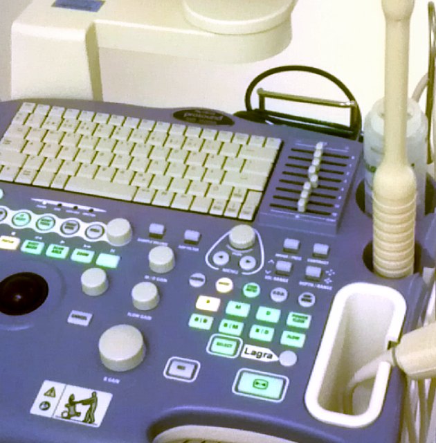
Ultrasound is the exam that locates the pregnancy. Beta-hCG helps interpret what the ultrasound shows and decide when to repeat it.
How accurate it is
The Brazilian review describes that transvaginal ultrasound identifies the ectopic with sensitivity of 87 to 94 per 100 and specificity of 94 to 99 per 100 when the exams are repeated (Pereira et al., 2019). With a single exam, sensitivity falls to 73.9 per 100, with specificity of 98.3 per 100.
This difference is the reason for follow-up. A first exam without findings does not end the investigation, and the repeated exam, with another beta-hCG value, often clarifies the picture.
The signs the examiner looks for
The 2025 review lists the main findings (Papageorgiou et al., 2025). The tubal ring sign, an echogenic ring image around the pregnancy outside the uterus, appears in 49 per 100 of ectopics and in 68 per 100 of unruptured ones, and when present it indicates tubal ectopic with a probability of 95 per 100.
Free fluid in the pelvis, added to a positive test and an empty uterus, had specificity of 70 per 100 and sensitivity of 63 per 100 (Papageorgiou et al., 2025). Fluid in the hepatorenal space is described as a strong marker of the need for surgery. Ring flow on Doppler can appear in the ectopic, but also in the corpus luteum, and the absence of flow does not exclude the diagnosis.

New locations, new names
A 2026 review draws attention to the rise in ectopics located inside the uterus but outside the cavity, especially in the cesarean scar (Farren et al., 2026). These pregnancies are harder to diagnose and to treat, and the review argues for new terminology to reduce the risk of error.
For clinical reasoning, the consequence is simple: "intrauterine pregnancy" in the report must say exactly where the sac is. A pregnancy implanted in the scar can look intrauterine at a quick glance.
Beta-hCG: from a single value to the curve
Discriminatory zone: the concept that lost strength
For a long time, the interpretation of ultrasound relied on a beta-hCG cutoff value. The idea seems logical, but studies have shown its limits.
What the discriminatory zone is
The discriminatory zone is the beta-hCG value above which an intrauterine gestational sac should be visible on ultrasound (Pereira et al., 2019). The Brazilian review describes that most services use values between 1500 and 2000 or 2500 mIU/mL and that, to have a 99 in 100 probability of seeing the sac, the value would need to reach 3510 mIU/mL.
The same review lists reasons for not seeing the sac even above the cutoff: multiple pregnancy, examiner experience, equipment resolution, obesity, fibroids, polyps and differences between laboratories (Pereira et al., 2019).
Why not use it to decide
The British review is more forceful. It states that the discriminatory zone should not be used to decide when to perform the ultrasound, because most ectopic pregnancies are seen with beta-hCG below 1000 IU/L, and that it is particularly important not to use the cutoff to presume ectopic pregnancy (Bobdiwala et al., 2017).
In practice, this means two things. A low beta-hCG is no reason to postpone the ultrasound for someone with symptoms. And a high beta-hCG with an empty uterus raises suspicion, but does not authorize treatment as ectopic without confirmation.
A high value also misleads
A British multicentre study analyzed 2507 women with a pregnancy of unknown location to test beta-hCG and progesterone cutoffs (Bobdiwala et al., 2022). No single beta-hCG value safely excluded a viable intrauterine pregnancy. Even above 3000 IU/L, 39 of 358 women, about 11 in 100, had a viable pregnancy.
Viable pregnancies appeared with progesterone as low as 5 nmol/L (Bobdiwala et al., 2022). The authors concluded that these cutoffs are not safe for excluding viability, and that a single beta-hCG used in this way is very unreliable.
The beta-hCG curve over 48 hours
If a single value misleads, the change over time tells you more. The standard is to compare two values collected about 48 hours apart.
Expected rise
The Brazilian review summarizes the change in criteria (Pereira et al., 2019). An older study showed that, in progressive intrauterine pregnancies with beta-hCG below 5000, the rise over 48 hours was at least 53 in 100. A more recent study with 1249 patients lowered that minimum to 35 in 100.
The 2023 review adds that a rise below 35 in 100 over 2 days suggests ectopic pregnancy, with accuracy of 80.2 in 100 (Mullany et al., 2023). Because accuracy is not perfect, the curve is one more piece of data for reasoning, not a verdict.
Fall and plateau
A fall in beta-hCG indicates a pregnancy that is not progressing. The Brazilian review describes that a ratio below 0.87 between the second and first value, that is, a fall greater than 13 in 100, suggests a failed pregnancy, with sensitivity of 92.7 in 100 and specificity of 96.7 in 100 (Pereira et al., 2019).
The plateau, with values that neither rise nor fall clearly, is the pattern that worries most. It does not confirm ectopic pregnancy, but it keeps the suspicion and calls for a new ultrasound and a new measurement.
| Beta-hCG pattern over 48 hours | What it suggests | What to do with the information |
|---|---|---|
| Rise of at least 35 in 100 | A progressive intrauterine pregnancy is possible | Repeat the ultrasound at the expected time to see the sac |
| Rise smaller than expected or plateau | Ectopic or failing pregnancy | Keep close follow-up, with ultrasound and a new measurement |
| Fall with ratio below 0.87 | Failed pregnancy | Follow until negative; ectopic is still possible, but less likely |
| High value with empty uterus | Greater suspicion of ectopic | Do not presume ectopic without confirmation; repeat and reassess |
Table source: Pereira et al., 2019; Mullany et al., 2023; Bobdiwala et al., 2017.
Beta-hCG and the risk of rupture
An Iranian study with 247 tubal ectopic pregnancies found rupture in 20.2 in 100 (Faraji Darkhaneh et al., 2015). Beta-hCG was higher in the ruptured cases, and values above 1750 were associated with rupture. Even so, ectopic pregnancies with low beta-hCG can also rupture, and the clinical picture remains paramount.
Risk models: who needs intensive follow-up
The idea behind the models is to use the trend in beta-hCG, sometimes together with progesterone, to estimate the risk of ectopic pregnancy and organize follow-up.
The two-step protocol
The British review describes a two-step protocol (Bobdiwala et al., 2017). First, the initial progesterone allows part of the women at very low risk to be discharged at the first visit. Then the others have a second beta-hCG drawn after 48 hours and are classified by a mathematical model.
Overall, the protocol classified 62.1 out of every 100 pregnancies of unknown location as low risk, with a negative predictive value of 98.6 out of every 100 and a sensitivity of 92.0 out of every 100 for classifying ectopic pregnancy as high risk (Bobdiwala et al., 2017).
Validation in other services
A Swedish study validated a model based only on two beta-hCG values in 1061 women seen in a gynecological emergency department, of whom 22 out of every 100 had an ectopic pregnancy (Fistouris et al., 2022). The area under the curve was 0.85 for the new model and 0.81 for the previous one.
With a threshold of 5 out of every 100 for high risk, the new model had a sensitivity of 95 out of every 100, a false-positive rate of 50 out of every 100 and a negative predictive value of 97 out of every 100 (Fistouris et al., 2022). In other words, it hardly lets any ectopic pregnancy slip through, at the cost of keeping many women who do not have one in follow-up.
A Pakistani study with 288 women, 22.9 out of every 100 with an ectopic pregnancy, compared three forms of triage (Izhar et al., 2022). The mathematical model had a sensitivity of 86.4 out of every 100 and a specificity of 91.9 out of every 100, while the criterion of progesterone alone had a specificity of only 27 out of every 100.
In practice, the lesson is that combining the information in a model yields more than looking at each number on its own, and that progesterone alone classifies poorly.
The case of assisted reproduction
In women who underwent in vitro fertilization, the date of transfer is known and beta-hCG is drawn on defined days. A Chinese study with 1703 women built a model with two hCG values and the cause of infertility to separate risk groups (Xu et al., 2020). This scenario shows how structured information improves risk estimation.
What the models do not do
The models do not give the diagnosis. They estimate the risk and help decide the intensity of follow-up. Clinical decision-making still depends on the clinical picture, the ultrasound and the conversation with the patient.
Instability and step-by-step reasoning
Instability: when reasoning changes pace
Everything said about follow-up applies to the stable patient. In the unstable patient, reasoning changes pace.
The signs that call for immediate action
Severe pain, signs of peritoneal irritation, fainting, tachycardia and hypotension in a woman with a positive test suggest a ruptured ectopic pregnancy until proven otherwise. The 2025 review notes that management depends on the integrity of the tube and on hemodynamic status, and that rupture requires surgery (Papageorgiou et al., 2025).
In this scenario, the 48-hour curve loses its meaning. Point-of-care ultrasound looks for free fluid, including in the hepatorenal space, and the surgical team is called in early (Papageorgiou et al., 2025).
When there is no ultrasound
The Tanzanian study showed that, without ultrasound, abdominal puncture was used for rapid diagnosis in part of the cases (Mooij et al., 2018). Women diagnosed by ultrasound received fewer transfusions and had less severe anemia than those diagnosed by puncture, and the authors highlight that physician delay in diagnosis was frequent.
Not every ectopic pregnancy needs intervention
At the opposite extreme, the 2026 review notes that better-quality ultrasound began detecting small ectopic pregnancies, many of them destined to regress on their own (Farren et al., 2026). The need to avoid overtreatment brought expectant management into clinical practice. Reasoning therefore ranges from the case that requires immediate surgery to the one that may only need monitoring.


Putting the reasoning together step by step
Suspected ectopic pregnancy can be organized in a sequence that respects uncertainty.
- Confirm the pregnancy with a test in every woman of reproductive age with abdominal pain or bleeding.
- Assess stability: vital signs, pain, peritoneal irritation.
- Unstable: bedside ultrasound looking for free fluid and call in the surgical team.
- Stable: transvaginal ultrasound, looking for an intrauterine sac and adnexal findings.
- No location: draw beta-hCG and repeat in about 48 hours.
- Use the curve and, if available, a risk model to decide the intensity of follow-up.
- Repeat the ultrasound as things evolve and explain each step and the warning signs to the patient.
Back to the patient in the case
The 29-year-old patient is stable, with an empty uterus and an intermediate beta-hCG. She has a pregnancy of unknown location, not a diagnosis of ectopic pregnancy (Pereira and colleagues, 2019). A single value does not rule out a viable pregnancy (Bobdiwala and colleagues, 2022), and an ultrasound with no findings does not end the investigation.
The next step is a new measurement in 48 hours and a new ultrasound as things evolve. If the curve rises adequately, an intrauterine pregnancy becomes more likely. If it falls, a failed pregnancy becomes more likely. If it plateaus, follow-up comes closer. In any case, she needs to leave knowing which signs require an immediate return, and the plan should be recorded in the medical record.
How NEXO helps train this reasoning
In NEXO, the case of the day is fictional, with a real clinical presentation and up to six progressive clues, and you propose the diagnosis by searching for the ICD-10 code.
The limits of using the game are described on the NEXO economy page. If you research the teaching of clinical reasoning, the page for researchers explains how to get in touch. Professionals who write or review cases find information on curation and on creators, and institutions can learn about the proposal on the partnerships page. Training differential diagnosis with clues and searching for the right ICD-10 code complete this training.
Frequently asked questions
Is an empty uterus with a positive test an ectopic pregnancy?
Not necessarily. It is a pregnancy of unknown location, which may end as an intrauterine pregnancy, a failed pregnancy or an ectopic pregnancy (Pereira and colleagues, 2019).
Is there a beta-hCG value that confirms or rules out an ectopic pregnancy?
No. The discriminatory zone should not be used to presume an ectopic pregnancy (Bobdiwala and colleagues, 2017), and even above 3000 IU/L about 11 in every 100 women had a viable pregnancy (Bobdiwala and colleagues, 2022).
How much should beta-hCG rise in 48 hours?
Recent studies indicate a minimum of about 35 in every 100 in progressive intrauterine pregnancies (Pereira and colleagues, 2019). A smaller rise or a plateau keeps the suspicion.
What are risk models for?
To organize follow-up. They identify who is at low risk and can have simplified follow-up and who needs intensive monitoring (Bobdiwala and colleagues, 2017).
Does every ectopic pregnancy need treatment?
No. Many small ectopic pregnancies regress on their own, and expectant management has entered practice to avoid excessive treatment (Farren and colleagues, 2026). The decision depends on stability and on how things evolve.
Take-home messages
- Pregnancy test in every woman of reproductive age with abdominal pain or bleeding.
- An empty uterus with a positive test is a pregnancy of unknown location.
- No single beta-hCG value rules out a viable pregnancy.
- The 48-hour curve tells you more than a single value.
- Risk models organize follow-up, but they do not replace ultrasound.
- Instability changes the pace: bedside ultrasound and early surgery.
References
- Bobdiwala S, Al-Memar M, Farren J, et al. Factors to consider in pregnancy of unknown location. Womens Health (Lond). 2017. PMID 28660799.
- Bobdiwala S, Kyriacou C, Christodoulou E, et al. Evaluating cut-off levels for progesterone, β human chorionic gonadotropin and β human chorionic gonadotropin ratio to exclude pregnancy viability in women with a pregnancy of unknown location: A prospective multicenter cohort study. Acta Obstet Gynecol Scand. 2022. PMID 34817062.
- Faraji Darkhaneh R, Asgharnia M, Farahmand Porkar N, et al. Predictive value of maternal serum β-hCG concentration in the ruptured tubal ectopic pregnancy. Iran J Reprod Med. 2015. PMID 25999999.
- Farren J, Al Wattar BH, Jurkovic D. The diagnosis and management of extrauterine and uterine ectopic pregnancy. Hum Reprod Update. 2026. PMID 41061761.
- Fistouris J, Bergh C, Strandell A. Pregnancy of unknown location: external validation of the hCG-based M6NP and M4 prediction models in an emergency gynaecology unit. BMJ Open. 2022. PMID 36446455.
- Izhar R, Husain S, Tahir MA, et al. Triaging Women with Pregnancy of Unknown Location: Evaluation of Protocols Based on Single Serum Progesterone, Serum hCG Ratios, and Model M4. J Reprod Infertil. 2022. PMID 36043136.
- Mooij R, Mgalega GC, Mwampagatwa IH, et al. A cohort of women with ectopic pregnancy: challenges in diagnosis and management in a rural hospital in a low-income country. BMC Pregnancy Childbirth. 2018. PMID 29751791.
- Mullany K, Minneci M, Monjazeb R, et al. Overview of ectopic pregnancy diagnosis, management, and innovation. Womens Health (Lond). 2023. PMID 36999281.
- Papageorgiou D, Sapantzoglou I, Prokopakis I, et al. Tubal Ectopic Pregnancy: From Diagnosis to Treatment. Biomedicines. 2025. PMID 40564186.
- Pereira PP, Cabar FR, Gomez ÚT, et al. Pregnancy of unknown location. Clinics (Sao Paulo). 2019. PMID 31618321.
- Wu JK, Sadecki EN, Kyweluk MA, et al. Patient attitudes and preferences for the management of pregnancy of unknown location. F S Rep. 2022. PMID 36212566.
- Xu H, Feng G, Wei Y, et al. Predicting Ectopic Pregnancy Using Human Chorionic Gonadotropin (hCG) Levels and Main Cause of Infertility in Women Undergoing Assisted Reproductive Treatment: Retrospective Observational Cohort Study. JMIR Med Inform. 2020. PMID 32297865.