Strongyloidiasis: hyperinfection in the immunosuppressed
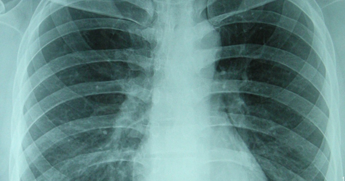
Table of contents
A 58-year-old woman, born in the interior of Bahia and living in São Paulo for decades, is admitted with severe pneumonia. She has been on corticosteroids for a few weeks for a rheumatologic disease. Within a few days, abdominal pain, diarrhea and bacteremia from an intestinal bacterium appear. The blood count shows no eosinophilia. The team treats the sepsis, but no one asks where she spent her childhood or thinks of a worm.
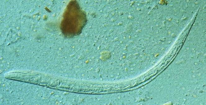
This is the most dangerous form of strongyloidiasis. Strongyloides stercoralis can live for decades in a person's intestine without causing almost anything. When immunity falls, mainly due to corticosteroids, the larval load explodes and the infection can spread through the body, carrying intestinal bacteria along with it. Without treatment, the course is usually fatal.
This text organizes the reasoning: why the parasite is different from the others, who is at risk, how to recognize hyperinfection when the classic signs are missing, which tests help and why the best strategy is to prevent before immunosuppressing.
Key points
- Strongyloides can multiply inside the host and persist for decades without symptoms.
- Corticosteroids are the most common trigger of hyperinfection, even in short courses.
- Eosinophilia is often absent precisely in severe cases.
- Sepsis or meningitis from an intestinal bacterium in an immunosuppressed patient should raise the suspicion.
- Parasitological tests and serology complement each other, and neither is perfect.
- Screening or treating those with exposure before immunosuppressing is the main measure.
Why this worm is different
A parasite that reproduces inside the host
Most helminths do not multiply inside the human body: to increase the load, the person needs to be reinfected. Strongyloides breaks this rule. Some of the larvae mature still in the intestine, cross the intestinal wall or the skin of the perianal region and restart the cycle, a process called autoinfection (Czeresnia and Weiss, 2022). That is why the infection can last a lifetime, even after the person has left the endemic area.
In most immunocompetent people, the chronic infection is asymptomatic or causes vague complaints, such as abdominal discomfort, intermittent diarrhea and pruritus. A more typical sign is larva currens, a linear, migratory skin lesion that advances quickly (Czeresnia and Weiss, 2022). These signs are subtle and easy to attribute to something else.
How many people carry the parasite
Estimates have changed a lot over the last decade. A 2013 review gathered 354 studies from 78 countries and showed that the disease is much more common than previously thought, with wide variation between regions (Schär et al., 2013). In 2020, a global model estimated a prevalence of 8.1 per 100, which corresponds to 613.9 million people infected, with 76.1 per 100 of them concentrated in Southeast Asia, Africa and the Western Pacific (Buonfrate et al., 2020). Another review speaks of more than 600 million infected (Czeresnia and Weiss, 2022).
Even these numbers may be below reality. The authors of the 2020 estimate note that many of the included studies used low-sensitivity tests, which leads to underestimation of prevalence, and that the disease is linked to each country's level of sanitation (Buonfrate et al., 2020).
Brazil stands out on this map. In the 2013 review, Brazil and Thailand were the countries with the most published studies, to the point of being shown separately in the graphs (Schär et al., 2013). This reflects both the presence of the parasite and a research tradition on the topic, and it reminds us that, here, strongyloidiasis is not a traveler's disease but a local infection.
Migration carries the parasite far from endemic areas. A prospective study in Catalonia, Spain, between 2003 and 2012, identified 70 patients with strongyloidiasis; 59 in 100 were considered to originate in South America and the Caribbean, 26 in sub-Saharan Africa, 13 in Southeast Asia and only 3 in 100 acquired locally (Czeresnia and Weiss, 2022). For the European physician, the Brazilian patient is one of the risk groups.
These numbers matter to the Brazilian physician. The parasite is transmitted by skin contact with contaminated soil, and tropical and subtropical regions, including rural areas of Brazil, are part of the risk map. The question about where the person was born, where they lived and whether they walked barefoot in childhood remains the first examination.
From silent carrier to hyperinfection
Hyperinfection is the acceleration of the autoinfection cycle, with a huge increase in the larval burden in the lungs and intestine. Disseminated disease is the migration of larvae to organs outside this axis, such as the central nervous system, liver and skin (Czeresnia and Weiss, 2022). In practice, the two terms overlap, and what changes management is recognizing that the infection has gone out of control.
The crucial point is that the larvae, as they cross the intestinal wall, carry bacteria from the flora. That is why hyperinfection often appears as bacteremia from enterobacteria or meningitis from intestinal germs, and not as an obvious parasitic disease (Czeresnia and Weiss, 2022). Without treatment, the reported mortality reaches 85 to 100 in 100.

Who is at risk
Corticosteroid, the most common trigger
The factor most associated with hyperinfection is systemic corticosteroid. In a review of 244 reports of hyperinfection and disseminated disease, 67 in 100 of the patients were using corticosteroids, almost always for another disease that already reduced immunity (Buonfrate and colleagues, 2013). Another review describes hyperinfection after short courses and at low doses, and recalls reports in patients who received corticosteroids for COVID-19 (Czeresnia and Weiss, 2022).
The lesson is uncomfortable: there is no known safe dose. The corticosteroid prescribed for an asthma exacerbation, for a flare of autoimmune disease or for a severe respiratory condition can be the trigger. The more common the use, the greater the number of people exposed to this risk.
HTLV-1 and transplantation
HTLV-1 is the second most recognized factor alongside corticosteroids (Siegel and Simon, 2012). In Okinawa, an endemic region for both agents, 36 of the 40 patients with disseminated disease who were tested had HTLV-1, or 90.0 in 100 (Mukaigawara and colleagues, 2020). This makes HTLV-1 serology a natural question in the face of severe strongyloidiasis, and vice versa.
Transplantation combines several risks: intense immunosuppression, corticosteroids and, sometimes, transmission through the organ itself. In a Brazilian study with 46 kidney transplant recipients with severe strongyloidiasis and 92 controls, the infection appeared at a median of 117 days after transplantation (Miglioli-Galvão and colleagues, 2020). The associated factors were deceased donor, with an odds ratio of 6.16, prior bacterial infection, with 3.04, and cumulative corticosteroid dose, with 1.005 per unit.
HIV, alcoholism and other factors
The relationship with HIV is less direct than was thought. The 2013 review found an odds ratio of 2.17 for Strongyloides infection in people with HIV and of 6.69 in people with alcoholism (Schär and colleagues, 2013). One proposed explanation is that HIV mainly compromises the Th1 response, while the Th2 response, which is more important against the worm, may be relatively preserved (Siegel and Simon, 2012).
The authors note that in advanced AIDS there is a profound loss of Th1 activity, but there may be little change in the cytokines of the Th2 response, such as interleukins 4, 5 and 13 (Siegel and Simon, 2012). The authors' conclusion is that HIV and corticosteroids, although seen as similar immunosuppressants, act in very different ways. In studies with transplant recipients, asthmatics, people with chronic lung disease and autoimmune diseases, corticosteroids were the common denominator of the severe forms (Siegel and Simon, 2012).
In practice, the risk list includes any relevant immunosuppression: chemotherapy, hematologic diseases, biologics and malnutrition. The reasoning is always the same: someone with old exposure to the parasite will lose part of their immunity, and that can wake up a forgotten infection.

How hyperinfection presents
The three Okinawa profiles
A series of 70 patients with disseminated strongyloidiasis admitted in Okinawa between 1975 and 2017 helped organize the clinical profiles (Mukaigawara and colleagues, 2020). The authors described three phenotypes: overt dissemination, in 45.7 per 100, occult dissemination with meningitis caused by gut bacteria, in 17.1, and occult dissemination with culture-negative purulent meningitis, in 37.1.
Severity varied widely between the groups. Mortality was 56.3 per 100 in overt dissemination, 16.7 in meningitis caused by gut bacteria and 11.5 in culture-negative meningitis. Sepsis was recorded in 40.6, 58.3 and 11.5 per 100, respectively. In total, 23 patients died, or 32.9 per 100.
The most common symptoms included fever, in 52.9 per 100, headache, in 32.9, and altered mental status, in 24.3 (Mukaigawara and colleagues, 2020). The authors stress that recognizing the occult form, milder and with striking neurological symptoms, saves lives.
Over the four decades of the series, patients were treated with thiabendazole until 2003 and with ivermectin after that (Mukaigawara and colleagues, 2020). For the clinician, the main message is different: faced with meningitis in an immunosuppressed patient from an endemic area, even with a negative culture, disseminated strongyloidiasis must be on the list.
What appears in transplant recipients
In the Brazilian study with kidney transplant recipients, gastrointestinal symptoms were present in 78.3 per 100, respiratory symptoms in 39.1 and fever in 32.6 (Miglioli-Galvão and colleagues, 2020). Eosinophilia appeared in 43.5 per 100, that is, it was absent in most. Mortality at 30 days was 28.3 per 100, reaching 47 when the infection occurred in the first three months after transplantation, against 17.2 after that.
The predictors of death were respiratory failure and bacteremia (Miglioli-Galvão and colleagues, 2020). This finding reinforces the clinical picture of hyperinfection: a pulmonary and abdominal presentation, often accompanied by severe bacterial infection.
Why eosinophilia misleads
Eosinophilia is the clue many expect, but it fails precisely in severe cases. Corticosteroids, which trigger hyperinfection, also reduce eosinophils, so the count is often normal (Czeresnia and Weiss, 2022). Even outside hyperinfection, in migrants from South Asia, 35 of the 115 who were seropositive for Strongyloides had no eosinophilia (Baker and colleagues, 2020).
A normal blood count, therefore, does not rule out the disease. Anyone who waits for eosinophilia to think of strongyloidiasis will miss exactly the patients who most need the diagnosis. The pretest probability comes from exposure and context, not from a single test.
Warning signs
Suspicion should arise when an immunosuppressed patient with possible exposure presents:
- bacteremia or meningitis caused by gut bacteria with no clear focus;
- cough, wheezing, chest pain, pneumonia or asthma that starts worsening again;
- abdominal pain, nausea, vomiting, diarrhea or ileus;
- larva currens or anal itching;
- clinical worsening shortly after starting or increasing corticosteroids.
This list follows the manifestations described in a recent review, which also includes invasion of organs such as the liver, pancreas and kidneys (Czeresnia and Weiss, 2022). Wheezing deserves special attention: a patient treated as severe asthma with corticosteroids may in fact be worsening the infection that causes the symptoms.
None of these signs is specific. What changes the reasoning is putting together the immunosuppression, the exposure history and an unexpected bacterial infection.
How to confirm the diagnosis
Parasitological tests
The shedding of larvae in stool is low and irregular in chronic infection, so a simple stool test often fails. The review of 143 studies on diagnostic methods concluded that direct microscopy is poorly sensitive (Requena-Méndez and colleagues, 2013). Specific methods, such as the Baermann method and agar plate culture, yield more: each has sensitivity of around 60 per 100, reaching 77 when combined (Carnino and colleagues, 2021).
In hyperinfection, the situation reverses. The larval load is so high that they can be seen in sputum, in bronchoalveolar or gastric lavage, in urine, in blood and even in biopsies of the lung or the gastrointestinal tract (Czeresnia and Weiss, 2022). The test depends, however, on someone remembering to order it. In the review of 244 reports, the diagnosis was made by microscopy in practically all of them, while serology was used in only 6.5 per 100 (Buonfrate and colleagues, 2013).
Serology
Serology is the most sensitive test for chronic infection, with sensitivity of 85 to 90 per 100 according to a review, lower in immunosuppressed patients (Carnino and colleagues, 2021). A study with 399 sera compared five tests and found sensitivity of 94.6 per 100 for an immunofluorescence assay and 92.3 for a commercial enzyme immunoassay, with specificity of 99.6 for the most specific test (Bisoffi and colleagues, 2014).
The study faced a problem common in the field: because the stool test is poorly sensitive, it is a weak reference standard. The authors therefore also used a composite standard, combining several tests, and observed that two commercial assays were highly specific above a given cutoff point (Bisoffi and colleagues, 2014). In practice, this means that the laboratory and the cutoff point used change the interpretation of the result.
Serology has limits. There can be cross-reaction with other helminths, and a positive result does not separate a current infection from an infection already cured (Requena-Méndez and colleagues, 2013). The review suggests repeating serology 6 to 12 months after treatment to follow the response.
Combining methods
Since no test is sufficient on its own, the recommendation is to combine direct and indirect methods (Carnino and colleagues, 2021). In screening those who will be immunosuppressed, serology is the best first step; when hyperinfection is suspected, looking for larvae in stool and respiratory secretions tends to yield more.
| Situation | Test that helps most | Main limit |
|---|---|---|
| Screening before immunosuppression | Serology | Cross-reaction; does not separate current from past infection |
| Chronic infection in an immunocompetent patient | Serology and concentration methods in stool | Low and irregular larval shedding |
| Suspected hyperinfection | Search for larvae in stool, sputum and lavage | Requires clinical suspicion to be ordered |
| Immunosuppressed patient with negative serology | Repeated parasitological methods | Serology less sensitive in immunosuppression |
| Follow-up after treatment | Follow-up serology | Titers fall slowly |


Prevent before immunosuppressing
Screening or presumptive treatment
The main measure against hyperinfection is to find and treat the infection before immunosuppression. A review recommends screening and treating those with exposure before starting corticosteroids or other immunosuppressants, and treating empirically when the result cannot come back in time (Czeresnia and Weiss, 2022). The same logic appears in a screening algorithm that combines exposure history, serology and parasitological methods (Carnino and colleagues, 2021).
The authors of this algorithm explain that many earlier proposals reflected the epidemiological context where they were created and could not be applied elsewhere (Carnino et al., 2021). Their proposal is based on two questions: what was the exposure and what will the intensity of immunosuppression be. The exposure assessment includes travel to rural areas, poor sanitation and the habit of walking barefoot, simple questions that fit into any consultation.
A systematic review concluded that serology is the most effective screening test in low-endemicity populations and that presumptive treatment with ivermectin is probably cost-effective (Agbata et al., 2018). A European economic study with migrants who would be immunosuppressed showed that presumptive treatment was a strategy that saved costs (Wikman-Jorgensen et al., 2020).
The screening that does not happen
The problem is that screening is rarely done. In the review of 244 reports, only 3 of the 29 transplant recipients had any mention of serological screening before transplantation (Buonfrate et al., 2013). The observed mortality was 68.5 per 100 in disseminated disease and 60 in hyperinfection, numbers that show the cost of this omission.
This finding has a simple reading for practice: the question about exposure needs to enter the checklist of anyone prescribing prolonged corticosteroids, chemotherapy, biologics or transplantation. Recording the decision in the medical record helps the team not to forget.
When the decision is urgent
There is not always time to wait for serology. The patient with severe disease who needs corticosteroids immediately cannot postpone treatment. In these cases, the review suggests empirical treatment when there is exposure and the test is not available in time (Czeresnia and Weiss, 2022). Weighing the risk of each path is the core of clinical decision-making, and the decision should be made with the infectious disease specialist when possible.
Mistakes that repeat
The first mistake is waiting for eosinophilia. The clue fails precisely in the patient using corticosteroids, and a normal blood count does not reduce the probability of the disease much.
The second mistake is treating the sepsis and not asking where the bacterium came from. Bacteremia or meningitis from a gut germ without a clear focus, in an immunosuppressed patient, should lead to a search for larvae.
The third mistake is forgetting old exposure. Someone born in an endemic rural area may carry the parasite for decades, even having lived in a big city for a long time.
The fourth mistake is trusting a single negative stool test. In chronic infection, larval shedding is irregular, and diagnostic uncertainty requires combining methods or repeating the test.
How NEXO helps train this reasoning
In NEXO, the case of the day is fictional, with a real clinical presentation and up to six progressive clues, and you propose the diagnosis by searching for the ICD-10 code.
The limits of using the game are described on the NEXO Economy page. If you research the teaching of clinical reasoning, the page for Researchers explains how to get in touch. Professionals who write or review cases find information under Curator and Creators, and institutions can learn about the proposal on the partnerships page. Training differential diagnosis with clues and searching for the right ICD-10 code complete this practice.
Frequently asked questions
Does a blood count without eosinophilia rule out strongyloidiasis?
No. Eosinophilia is often absent in hyperinfection, because the corticosteroid that triggers the condition also reduces eosinophils (Czeresnia and Weiss, 2022). In the Brazilian study with transplant recipients, it appeared in 43.5 per 100 (Miglioli-Galvão et al., 2020).
Who should be screened before receiving corticosteroids?
People with possible exposure, such as those born or who lived in a tropical rural area, and who will receive corticosteroids or other immunosuppression. Serology is the most indicated screening test (Agbata et al., 2018).
Why think of a worm when facing sepsis?
Because the larvae carry intestinal bacteria as they cross the intestinal wall. Bacteremia and meningitis from enterobacteria are common forms of presentation of hyperinfection (Czeresnia and Weiss, 2022).
Does a positive serology mean active infection?
Not necessarily. Serology does not separate current infection from past infection and may cross-react with other helminths (Requena-Méndez et al., 2013).
What is the relationship with HTLV-1?
HTLV-1 is one of the most recognized factors for severe forms (Siegel and Simon, 2012). In Okinawa, 90.0 out of every 100 patients tested with disseminated disease had the virus (Mukaigawara et al., 2020).
Take-home messages
- Strongyloides multiplies inside the host and can persist for decades.
- Corticosteroids are the main trigger of hyperinfection.
- Absent eosinophilia does not rule out the disease, especially in the immunosuppressed.
- Sepsis or meningitis caused by a gut organism should raise suspicion.
- Serology for screening; larval testing when hyperinfection is suspected.
- Screening or treating before immunosuppression is the measure that saves the most lives.
References
- Agbata EN, Morton RL, Bisoffi Z, et al. Effectiveness of Screening and Treatment Approaches for Schistosomiasis and Strongyloidiasis in Newly-Arrived Migrants from Endemic Countries in the EU/EEA: A Systematic Review. Int J Environ Res Public Health. 2018. PMID 30577567.
- Baker EC, Ming DK, Choudhury Y, et al. High Prevalence of <i>Strongyloides</i> among South Asian Migrants in Primary Care-Associations with Eosinophilia and Gastrointestinal Symptoms. Pathogens. 2020. PMID 32041352.
- Bisoffi Z, Buonfrate D, Sequi M, et al. Diagnostic accuracy of five serologic tests for Strongyloides stercoralis infection. PLoS Negl Trop Dis. 2014. PMID 24427320.
- Buonfrate D, Bisanzio D, Giorli G, et al. The Global Prevalence of <i>Strongyloides stercoralis</i> Infection. Pathogens. 2020. PMID 32545787.
- Buonfrate D, Requena-Mendez A, Angheben A, et al. Severe strongyloidiasis: a systematic review of case reports. BMC Infect Dis. 2013. PMID 23394259.
- Carnino L, Schwob JM, Gétaz L, et al. A Practical Approach to Screening for <i>Strongyloides stercoralis</i>. Trop Med Infect Dis. 2021. PMID 34941659.
- Czeresnia JM, Weiss LM. Strongyloides stercoralis. Lung. 2022. PMID 35396957.
- Miglioli-Galvão L, Pestana JOM, Santoro-Lopes G, et al. Severe Strongyloides stercoralis infection in kidney transplant recipients: A multicenter case-control study. PLoS Negl Trop Dis. 2020. PMID 32004346.
- Mukaigawara M, Narita M, Shiiki S, et al. Clinical Characteristics of Disseminated Strongyloidiasis, Japan, 1975-2017. Emerg Infect Dis. 2020. PMID 32091375.
- Requena-Méndez A, Chiodini P, Bisoffi Z, et al. The laboratory diagnosis and follow up of strongyloidiasis: a systematic review. PLoS Negl Trop Dis. 2013. PMID 23350004.
- Schär F, Trostdorf U, Giardina F, et al. Strongyloides stercoralis: Global Distribution and Risk Factors. PLoS Negl Trop Dis. 2013. PMID 23875033.
- Siegel MO, Simon GL. Is human immunodeficiency virus infection a risk factor for Strongyloides stercoralis hyperinfection and dissemination. PLoS Negl Trop Dis. 2012. PMID 22860137.
- Wikman-Jorgensen PE, Llenas-Garcia J, Shedrawy J, et al. Cost-effectiveness of different strategies for screening and treatment of <i>Strongyloides stercoralis</i> in migrants from endemic countries to the European Union. BMJ Glob Health. 2020. PMID 32461226.