Athlete ECG: physiological adaptation or disease



Table of contents
- Why the athlete's ECG is different
- The evolution of interpretation criteria
- What current criteria consider normal, borderline and abnormal
- The T wave: the finding that demands the most care
- Ancestry, age and sex change the reading
- What the ECG does not see
- From the abnormal finding to the decision
- How NEXO helps train this reasoning
- Frequently asked questions
- Take-home messages
- References
A 19-year-old basketball player has an ECG as part of preseason screening. The tracing shows a rate of 46 beats per minute, incomplete right bundle branch block and tall QRS complexes in the chest leads. The physician reading the exam must decide whether all of this is the trained heart working as it should or whether there is a cardiomyopathy hidden behind findings that look benign.
This decision is the core of sports cardiology. Intense, repeated training changes the heart's electrical activity and structure, and many of these changes mimic disease patterns (Basu, 2018). Misreading the ECG causes harm on both sides: reassuring someone who has disease, or removing a healthy person from sport and subjecting them to tests.
This text shows how interpretation criteria evolved, what is now considered normal, borderline and abnormal, and where reading becomes harder: adolescents, Black athletes and athletes over 35.
Key points
- The 2005 criteria classified the ECG of up to half of athletes as abnormal, and later revisions greatly reduced false positives (Basu, 2018).
- The 2017 international criteria separate normal training findings, borderline findings and abnormal findings; two or more borderline findings together become abnormal (Schneiter, 2018).
- T wave inversion beyond V2 is rare after puberty in white athletes and calls for investigation (Pieles, 2026).
- In Black athletes, T wave inversion from V1 to V4 preceded by convex ST elevation is considered a normal variant (Basu, 2018).
- Under 16, T wave inversion from V1 to V3 is part of the juvenile pattern (Pieles, 2026).
- Over 35, coronary disease becomes the main target, and the criteria designed for young athletes perform less well (Panhuyzen-Goedkoop, 2020).
Summary
- Why the athlete's ECG is different
- The evolution of interpretation criteria
- What current criteria consider normal, borderline and abnormal
- The T wave: the finding that demands the most care
- Ancestry, age and sex change the reading
- What the ECG does not see
- From abnormal finding to decision
- How NEXO helps train this reasoning
- Frequently asked questions
- Take-home messages
Why the athlete's ECG is different
The heart of someone who trains many hours a week adapts. Vagal tone increases and produces sinus bradycardia, sinus arrhythmia, first-degree atrioventricular block and even second-degree Mobitz I block (Basu, 2018). The chambers enlarge and the left ventricular wall becomes thicker, which raises QRS voltage.
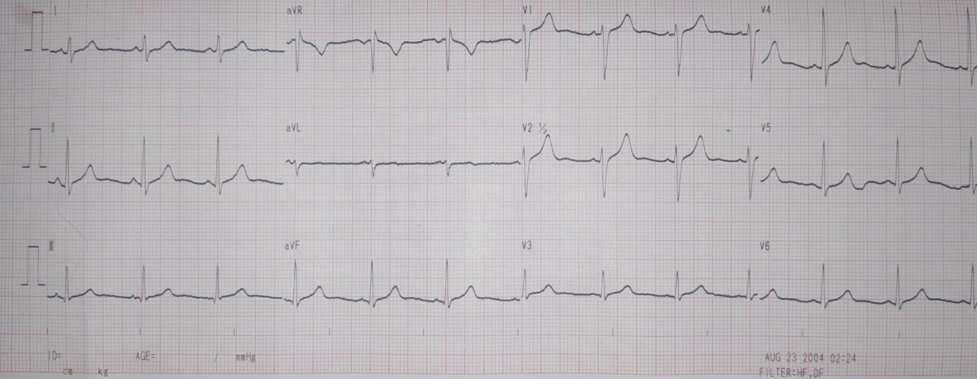
These changes are frequent. Incomplete right bundle branch block, with QRS under 120 milliseconds, appears in 30 to 50 of every 100 athletes (Basu, 2018). Early repolarization, with J point elevation, is also common and is considered normal in all current criteria (Halasz, 2025).
The problem is that the diseases that most often kill young athletes also change the ECG. Up to 95 of every 100 people with hypertrophic cardiomyopathy and about 80 of every 100 with arrhythmogenic right ventricular cardiomyopathy have an abnormal ECG (Basu, 2018). The ECG is also the exam that detects electrical syndromes such as long QT, Brugada and Wolff-Parkinson-White.
The task, therefore, is to separate what training explains from what it does not. The logic is the same as in any differential diagnosis: each finding changes the probability of disease, and some change it far more than others.
What is being looked for: the causes of sudden death in young athletes
Knowing what is being looked for helps in reading the tracing. In a four-year American prospective registry, the most common causes of cardiac arrest or sudden death in young athletes were hypertrophic cardiomyopathy, idiopathic left ventricular hypertrophy, coronary artery anomalies and sudden death with a normal autopsy (Peterson, 2021). The athletes' mean age was 16.7 years, and most were male and played basketball or American football (Peterson, 2021).
The profile changed with age. Coronary anomalies were more frequent among middle school athletes, while cardiomyopathies accounted for almost half of events among college and professional athletes (Peterson, 2021). This detail matters because cardiomyopathies usually change the resting ECG, and coronary anomalies do not.
The incidence was also higher in men. In high school, the rate ratio between men and women was 4.6 (Peterson, 2021). The authors conclude that ECG screening may be more effective in higher-risk groups of athletes.
The evolution of interpretation criteria
The first criteria for the athlete's ECG were published by the European Society of Cardiology in 2005. With them, the ECG was classified as abnormal in up to half of athletes, a false-positive rate that is unacceptable for a screening program (Basu, 2018).
In 2010, the same society separated training-related changes from non-training-related ones. In 2013 came the Seattle criteria and, in 2014, the so-called refined criteria, which began to treat some findings previously considered abnormal as borderline, such as isolated axis deviation and atrial overload (Basu, 2018). The sequence culminated in the 2017 international recommendations, which reduced false positives and the cost of additional investigation.
The effect of these revisions was measured directly. In a study of 2491 Arab, Black and White male athletes, the proportion of abnormal ECGs fell from 22.3 per 100 with the 2010 European criteria to 11.6 with Seattle and 5.3 with the refined criteria (Riding, 2015). All three criteria identified every athlete with hypertrophic cardiomyopathy and Wolff-Parkinson-White, and specificity rose to 94.0 per 100 with the refined criteria (Riding, 2015).
Two changes accounted for most of the gain: higher cutoffs for QTc, 470 milliseconds in men and 480 in women, and the recognition that isolated anterior T-wave inversion in asymptomatic Black athletes is benign (Riding, 2015).
What current criteria consider normal, borderline and abnormal
The international criteria organize findings into three groups (Halasz, 2025; Schneiter, 2018). The first gathers the normal adaptations to training, which need no investigation. The second gathers borderline findings, acceptable when isolated. The third gathers changes not explained by training, which require evaluation.
Normal findings of the trained heart
Sinus bradycardia, sinus arrhythmia, first-degree atrioventricular block, incomplete right bundle branch block, early repolarization and isolated increase in QRS voltage are considered normal adaptations (Halasz, 2025). The voltage increase deserves a comment: in hypertrophic cardiomyopathy, it usually comes with T-wave inversion, Q waves or ST depression (Basu, 2018). On its own, it is a sign of training.
Borderline findings
Left or right axis deviation, left or right atrial overload and complete right bundle branch block belong in this group (Corsi, 2025). A study of more than 2000 athletes and nearly 10,000 controls found no relevant structural or functional changes in those with isolated axis deviation or atrial overload (Basu, 2018).
The practical rule is that of the sum. An isolated borderline finding is treated as normal; two or more together are considered abnormal and require investigation (Schneiter, 2018).
Abnormal findings
This group includes T-wave inversion outside accepted patterns, ST-segment depression, pathological Q waves, ventricular pre-excitation and prolonged QTc above the limits (Halasz, 2025). The European sports cardiology guideline notes that extensive T-wave inversion, in three or more leads from V1 to V6, or in two of the three inferior leads, carries additional risk of sudden death (Pelliccia, 2020).
| Group | Examples of findings | What to do | Source |
|---|---|---|---|
| Normal for training | Sinus bradycardia, incomplete right bundle branch block, early repolarization, isolated QRS voltage | No investigation needed in the absence of symptoms | Halasz, 2025 |
| Borderline | Axis deviation, atrial overload, complete right bundle branch block | Acceptable when isolated; two or more together require investigation | Schneiter, 2018 |
| Abnormal | T-wave inversion outside the accepted pattern, ST depression, pathological Q waves, pre-excitation, long QTc | Investigation before clearing for competition | Halasz, 2025 |
| Ethnic variant | T-wave inversion from V1 to V4 with convex ST elevation in Black athletes | Considered normal when isolated and without symptoms | Basu, 2018 |
| Juvenile pattern | T-wave inversion from V1 to V3 before age 16 | Considered normal for age | Pieles, 2026 |
The T wave: the finding that demands the most care
T-wave inversion is where adaptation and disease are most easily confused. Location matters a great deal. In a study described in Basu's review, anterior T-wave inversion, from V1 to V4, appeared in 12.7 out of every 100 Black athletes and was not associated with disease, while lateral inversion was (Basu, 2018).
In White athletes who have completed puberty, anterior T-wave inversion beyond V2 is rare, present in about 0.1 out of every 100 (Pieles, 2026). For this reason, it is treated as abnormal in most adults. Lateral or inferolateral inversion, in any athlete, is one of the findings that most calls for investigation, because it can be the first sign of cardiomyopathy (Pelliccia, 2020).
For the player in the opening scene, this means looking carefully at repolarization. Bradycardia, incomplete right bundle branch block and high voltage, without out-of-pattern T-wave inversion, without Q waves and without ST depression, form a set typical of the trained heart (Halasz, 2025). If the same player had T-wave inversion from V4 to V6, the conversation would change.
Ancestry, age and sex change the reading
Black athletes
The hearts of Black athletes tend to show more pronounced adaptations. T-wave inversion from V1 to V4 preceded by convex ST elevation came to be considered a normal ethnic variant (Basu, 2018), and this change was one of the main reasons for the drop in false positives (Riding, 2015).
A 2025 systematic review of 47 studies questions the use of broad racial categories (Corsi, 2025). The frequency of the so-called Black athlete repolarization variant ranged from 1.8 per 100 in Brazilians to 30 per 100 in Ghanaians, and T-wave inversion varied up to fourfold within Black populations (Corsi, 2025). Even with current criteria, the false-positive rate was 3.3 per 100 in Black athletes and 1.4 per 100 in White athletes (Corsi, 2025). The authors suggest that geographic ancestry explains adaptations better than skin color.
Adolescents
Before puberty, anterior T-wave inversion is common and part of the juvenile pattern, because of right ventricular dominance. The European document on pediatric athletes considers inversion from V1 to V3 before age 16 and from V1 to V4 before age 12 to be normal (Pieles, 2026). Many findings of the trained adult, such as sinus bradycardia, incomplete right bundle branch block and early repolarization, also appear in young athletes.
The same document recommends repeating the assessment every two years until age 16 and using the adult documents from that age on (Pieles, 2026).
Master athletes
Above age 35, the target changes. In a Dutch study of 2578 master athletes, coronary disease was the most frequently detected high-risk condition (Panhuyzen-Goedkoop, 2020). The older criteria detected more high-risk conditions than the international ones, because the latter treat isolated atrial overload and voltage as normal or borderline.
In an Italian study of 506 master athletes, 13 had a condition associated with sudden death, and the Seattle criteria performed better than the international ones (Halasz, 2025). False-positive rates were 30.4 per 100 with the European criteria, 21.9 with Seattle and 15 with the international ones (Halasz, 2025). The authors suggest that master athletes call for their own criteria, which give more weight to axis deviation and family history.
The most common findings in master athletes
In the Italian study, the most frequent abnormal findings in master athletes were left axis deviation, left atrial overload and T-wave inversion (Halasz, 2025). The international criteria failed to identify two athletes with dilated cardiomyopathy who had left axis deviation, precisely because this finding, in isolation, is treated as borderline.
Complete right bundle branch block, on the other hand, was not associated with relevant structural changes in these athletes (Halasz, 2025). And the diagnosis of long QT varied widely depending on the criterion used, which shows how cutoffs influence the number of athletes referred for investigation.
In practice, the master athlete calls for an assessment that goes beyond the ECG, with attention to cardiovascular risk factors and the history of symptoms on exertion (Panhuyzen-Goedkoop, 2020). The reasoning is close to the one used for non-athlete adults with coronary risk.
What the ECG does not see
A normal ECG lowers the probability of some diseases, but not all of them. Coronary artery anomalies usually have a normal ECG and present as chest pain or syncope during exertion (Peterson, 2021). In an American registry of 331 cardiac arrest or sudden death events in young athletes, coronary anomalies accounted for a relevant share of the events, especially among the youngest.
That is why symptoms during exertion still count for more than a normal tracing. Syncope on exertion, in particular, calls for cardiological investigation regardless of the resting ECG, as in the assessment of signs that point to a cardiac cause in syncope in general.
At the same time, the registry showed that 71 of every 100 events with a defined cause in college and professional athletes were caused by diseases that usually alter the ECG (Peterson, 2021). A meta-analysis of 28,011 athletes aged 10 to 35 found a much stronger association between the ECG and the detection of conditions linked to sudden death than between history and physical examination and that detection (Goff, 2023).
Who reads the ECG also makes a difference
Standardized criteria only work if they are applied consistently. In a Swiss study, a medical student, a cardiology resident and an electrophysiologist independently read 287 ECGs of elite athletes using the international criteria (Schneiter, 2018). Agreement for abnormal findings was excellent, with kappa between 0.855 and 1.000, and overall agreement was moderate to good (Schneiter, 2018).
The result suggests that clear criteria reduce dependence on experience for the most important findings. Disagreements were concentrated in normal and borderline findings, precisely where the decision has fewer consequences (Schneiter, 2018).
Anyone who reads athletes' ECGs also needs to know the pitfalls common to any tracing. Metabolic changes, such as those described in the text on hyperkalemia on the ECG, and electrode placement errors can mimic abnormal findings.
From the abnormal finding to the decision
An abnormal ECG is not a diagnosis; it is the start of an investigation. For abnormal T wave inversion, the European document on young athletes suggests echocardiography, an exercise test and 24-hour Holter monitoring, with cardiac magnetic resonance when there is suspicion or abnormal findings, and genetic evaluation if there are signs of disease or a family history (Pieles, 2026).
The pretest probability guides the intensity of the investigation. A lateral T wave inversion in an athlete with a family history of sudden death means something very different from an isolated axis deviation in an athlete without symptoms, reasoning detailed in the text on pretest probability. The same numbers of sensitivity and predictive values explain why the false positive rate depends so much on the population tested.
Excess must also be avoided. Each false positive generates tests, anxiety and sometimes undue removal from sport (Basu, 2018). Cascades of care start like this, with a borderline finding read as abnormal. When the investigation does not clarify the picture, serial reassessment may be more useful than definitive removal, as in watchful waiting with a safety net.
The decision about sport, when there is confirmed disease, is individual and shared with the athlete, according to the European guideline (Pelliccia, 2020). The same logic of assessing the risk of a sudden event appears in other contexts, such as the one-in-a-hundred-per-year rule used in aviation.
A reading script for the player in the scene
Back to the 19-year-old player: the reading can follow a fixed order. First, confirm the quality of the tracing and the position of the electrodes. Second, list the findings and classify each one as normal for training, borderline or abnormal, according to the international criteria (Schneiter, 2018). Third, count the borderline findings: one alone does not call for investigation, two or more do.
Fourth, look at repolarization lead by lead, remembering that age, ancestry and location change the meaning of the T wave (Pieles, 2026). Fifth, go back to the history: symptoms on exertion, family history of sudden death and medication use. A typical tracing of a trained heart, in an athlete with no symptoms and no family history, allows clearance with confidence.
If any of these steps raises doubt, the investigation follows the logic of the finding, and the result must be explained to the athlete clearly. The European guideline encourages shared decision-making and respect for the athlete's autonomy after detailed information about risks (Pelliccia, 2020).
How NEXO helps train this reasoning
In NEXO, the case of the day is fictional, with a real clinical presentation and up to six progressive clues, and you propose the diagnosis by searching for the ICD-10 code.
The limits on using the game are described on the NEXO Economy page. If you research the teaching of clinical reasoning, the Researcher page explains how to get in touch. Professionals who write or review cases find information on the Curator and Creators pages, and institutions can learn about the proposal on the partnerships page. To practice finding the code, see how the ICD-10 code is built.
Frequently asked questions
Is bradycardia in the athlete always normal?
Sinus bradycardia is a common adaptation to training and is considered normal in current criteria (Halasz, 2025). Symptoms such as syncope or dizziness on exertion change the assessment.
Does right bundle branch block keep the athlete away from sport?
Incomplete block is normal and appears in 30 to 50 out of every 100 athletes (Basu, 2018). Complete block is borderline and only calls for investigation if it comes with another borderline finding.
Does high QRS voltage mean diseased hypertrophy?
On its own, it is a sign of training. In hypertrophic cardiomyopathy, it usually comes with T wave inversion, Q waves or ST depression (Basu, 2018).
Is T wave inversion always disease?
It depends on age, ancestry and lead. Before age 16, inversion in V1 to V3 is a juvenile pattern (Pieles, 2026); lateral inversions call for investigation in any athlete (Pelliccia, 2020).
Do the criteria for young people apply to athletes over 35?
They apply with limitations. In master athletes, performance drops and coronary disease becomes the main target (Halasz, 2025; Panhuyzen-Goedkoop, 2020).
Does a normal ECG guarantee that the athlete has no heart disease?
It guarantees little for coronary anomalies, which usually have a normal ECG (Peterson, 2021). Symptoms on exertion always call for investigation.
Take-home messages
- Training produces bradycardia, incomplete right bundle branch block, early repolarization and high voltage, which are normal in the athlete.
- Axis deviation, atrial overload and complete right bundle branch block are borderline; two or more together call for investigation.
- Lateral T wave inversion, ST depression, pathological Q waves, pre-excitation and long QTc are abnormal.
- Reading the T wave depends on age and ancestry, and broad racial categories generate false positives.
- Above age 35, the focus shifts to coronary disease and the criteria for young people perform less well.
- Symptoms on exertion count for more than a normal ECG, because some diseases do not change the tracing.
- An abnormal ECG opens an investigation; the decision about sport is individual and shared.
Play the case of the day on NEXO.
References
- Basu J, Malhotra A. Interpreting the Athlete's ECG: Current State and Future Perspectives. Curr Treat Options Cardiovasc Med. 2018. PMID 30456469. DOI 10.1007/s11936-018-0693-0.
- Riding NR, Sheikh N, Adamuz C, et al. Comparison of three current sets of electrocardiographic interpretation criteria for use in screening athletes. Heart. 2015. PMID 25502812. DOI 10.1136/heartjnl-2014-306437.
- Schneiter S, Trachsel LD, Perrin T, et al. Inter-observer agreement in athletes ECG interpretation using the recent international recommendations for ECG interpretation in athletes among observers with different levels of expertise. PLoS One. 2018. PMID 30462649. DOI 10.1371/journal.pone.0206072.
- Halasz G, Capelli B, Crispino SP, et al. The diagnostic accuracy of contemporary ECG interpretation criteria in risk stratification of master athletes. Front Cardiovasc Med. 2025. PMID 40964528. DOI 10.3389/fcvm.2025.1637853.
- Panhuyzen-Goedkoop NM, Wellens HJ, Verbeek AL, et al. ECG criteria for the detection of high-risk cardiovascular conditions in master athletes. Eur J Prev Cardiol. 2020. PMID 31996014. DOI 10.1177/2047487319901060.
- Corsi D, Hernandez R, Bao JY, et al. Beyond Racial Categorization in Sports Cardiology: A Systematic Review of Cardiac Adaptations in Athletes. J Clin Med. 2025. PMID 41096186. DOI 10.3390/jcm14197107.
- Pieles GE, Cavarretta E, Orchard JJ, et al. Cardiac evaluation of paediatric athletes. Eur Heart J. 2026. PMID 41967042. DOI 10.1093/eurheartj/ehag188.
- Pelliccia A, Sharma S, Gati S, et al. 2020 ESC Guidelines on sports cardiology and exercise in patients with cardiovascular disease. Eur Heart J. 2021. PMID 32860412. DOI 10.1093/eurheartj/ehaa605.
- Peterson DF, Kucera K, Thomas LC, et al. Aetiology and incidence of sudden cardiac arrest and death in young competitive athletes in the USA: a 4-year prospective study. Br J Sports Med. 2021. PMID 33184114. DOI 10.1136/bjsports-2020-102666.
- Goff NK, Hutchinson A, Koek W, et al. Meta-analysis on the Effectiveness of ECG Screening for Conditions Related to Sudden Cardiac Death in Young Athletes. Clin Pediatr (Phila). 2023. PMID 36797841. DOI 10.1177/00099228231152857.